


 下载:
下载:
-
骨岛又被称为骨斑或内生骨疣,是位于松质骨内大小不一的致密骨,好发于骨盆、股骨及其他长骨,一般是由软骨内骨化过程中的异常引起[1]。常见的骨岛多较小,大部分患者无临床症状,骨扫描通常未见代谢增高[2],临床上显示为代谢增高的骨岛较少见[3-6],易造成误诊。我们分析了1例最终经组织病理学检查结果证实的99Tcm-MDP SPECT/CT全身骨显像代谢增高的骨岛患者的影像表现特点及鉴别诊断过程,以提高影像医师对该病的认识。
-
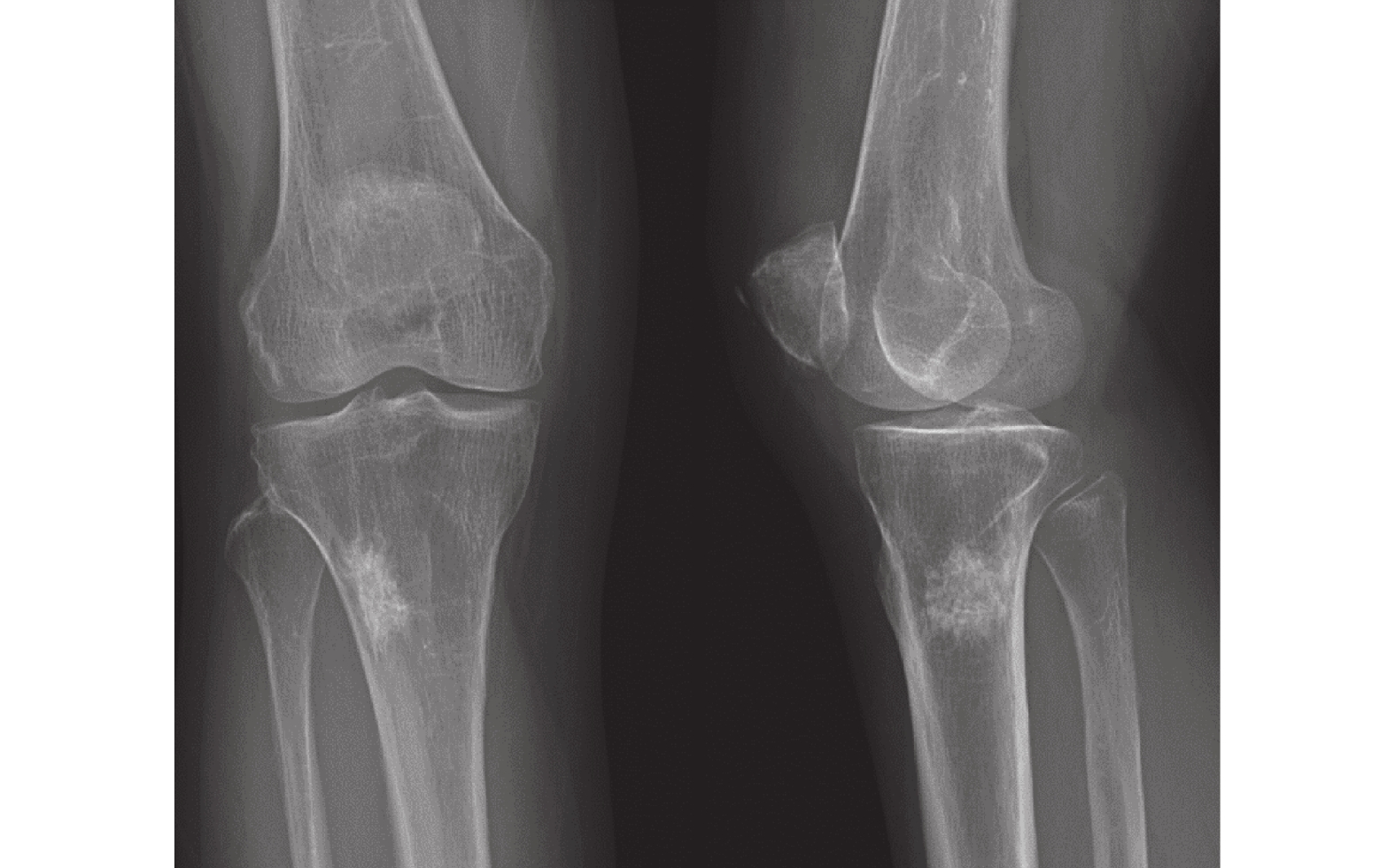
患者女性,66岁,因“右膝部疼痛1个月,加重10 d”就诊。患者1个月前出现无明显诱因右侧膝部疼痛,间歇性发作,活动及下蹲后加重,10 d前患者症状加重,遂就诊于四川省肿瘤医院。2年前,患者因盆腔肿块行手术治疗,术后组织病理学检查结果示阑尾低级别黏液性肿瘤。体格检查结果示右膝部外观无异常,未扪及肿块,皮温正常,局部无红肿,皮肤无破溃。实验室检查结果示血尿便常规、肝肾功未见明显异常。膝部X线正侧位片示右侧胫骨上段不规则片状致密影,大小约2.4 cm×1.3 cm×3.0 cm,边缘呈锯齿状,周围骨皮质无骨膜反应(图1)。
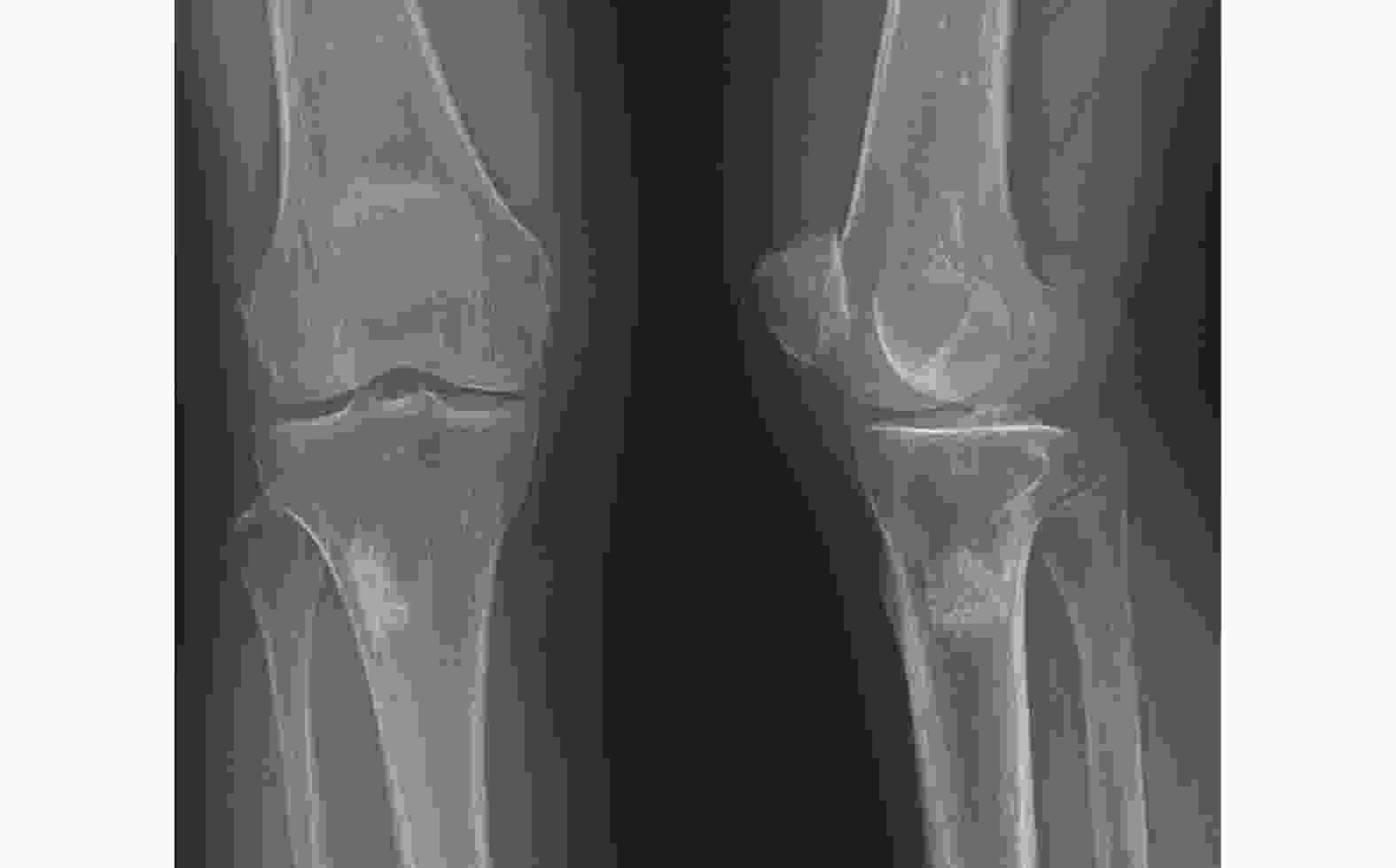
图 1 右胫骨骨岛患者(女性,66岁)的膝部X线正侧位图
Figure 1. Anteroposterior radiograph of the knee of a patient (female, 66 years old) with bone island of right tibia
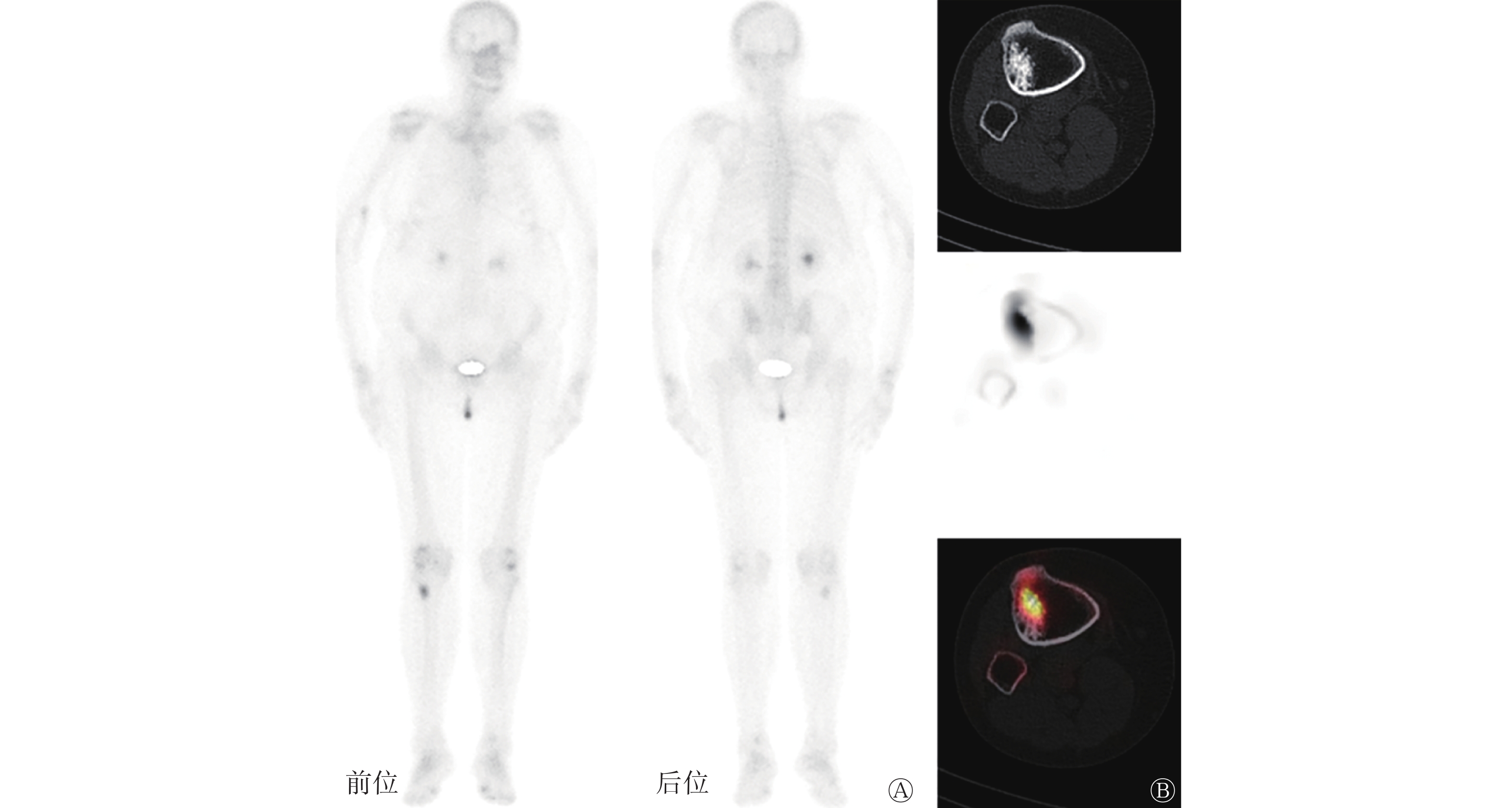
因患者有既往肿瘤病史,为进一步评估病情,行99Tcm-MDP全身骨显像,结果示右侧胫骨上段有代谢增高灶,余全身骨骼未见明显代谢异常征象;遂加行右胫骨上段SPECT/CT融合显像,相应层面CT、SPECT、SPECT/CT图示右侧胫骨上段有不规则团块状致密影,周围见“伪足”,伴异常放射性浓聚(图2),考虑为骨岛、内生软骨瘤或成骨转移。
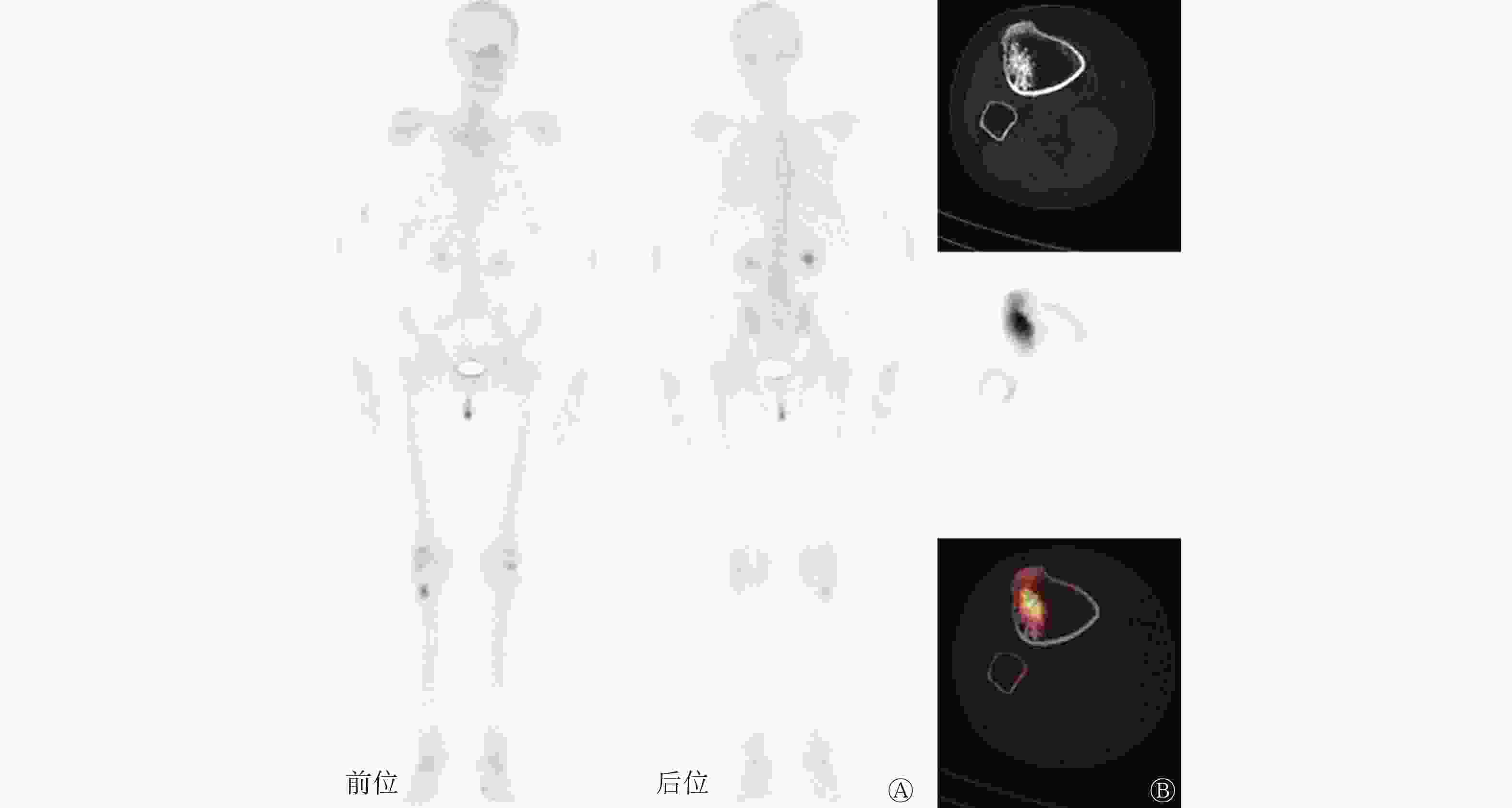
图 2 右胫骨骨岛患者(女性,66岁)的99Tcm-MDP SPECT/CT全身骨显像图
Figure 2. 99Tcm-MDP SPECT/CT whole-body bone imaging of a patient (female, 66 years old) with bone island of right tibia
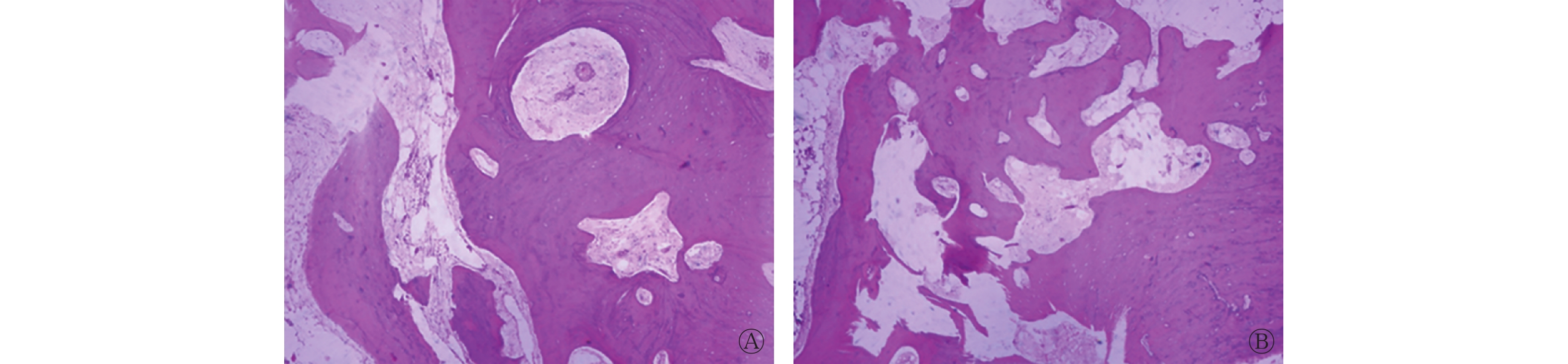
由于患者疼痛症状明显,要求手术治疗,遂于全麻下行“右胫骨骨肿瘤切除瘤腔灭活术”,术中见右胫骨上段骨膜无明显水肿或增厚,骨皮质外观无异常,病灶骨皮质内侧为质硬灰样物质,髓腔无明显异常,未见确切软组织。术后组织病理学检查结果示髓内位置为正常哈弗氏管及不规则成熟板层骨,无有丝分裂或非典型细胞核(图3),提示该病灶为骨岛。术后患者疼痛缓解。
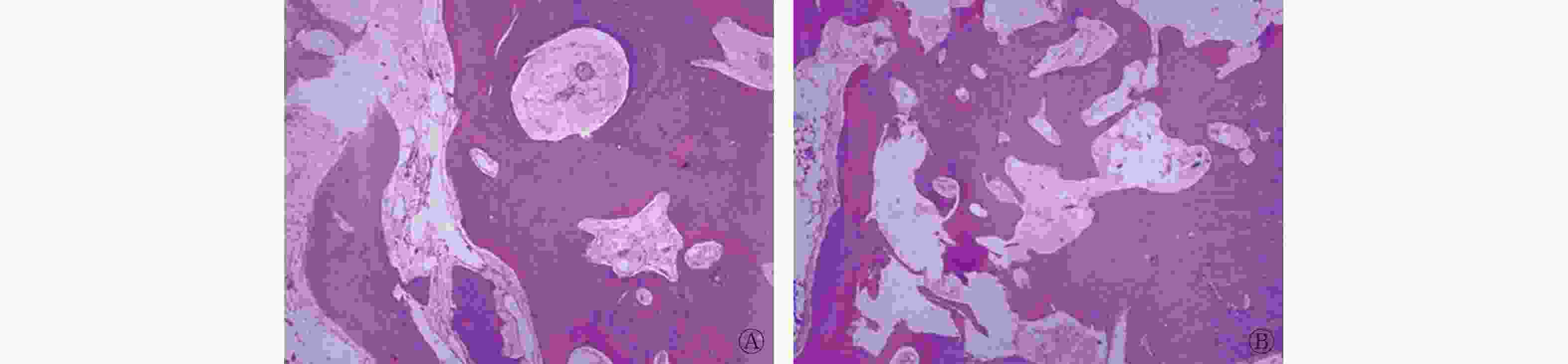
图 3 右胫骨骨岛患者(女,66岁)的术后组织病理学检查图(苏木精-伊红染色,×40,脱钙切片)
Figure 3. Postoperative histopathological examination of a patient (female, 66 years old) with bone island of right tibia
-
骨岛是位于松质骨内大小不一的致密骨[1]。它是一种稳定的病变,形状和大小多无明显变化,大多数的骨岛都比较小,通常<2 cm,而>2 cm者称为巨大骨岛[7]。骨岛生长方向通常与骨长轴一致,组织病理学表现为成熟的板层骨及完整的哈弗氏系统,多由硬骨板和增厚的骨小梁组成,无成骨细胞。大部分骨岛患者无临床症状,多为X线或CT检查时偶然发现。骨岛常有较为典型的影像表现,在X线片及CT图像上表现为松质骨内圆形或类圆形骨样高密度影,部分周围见“伪足”,与周边骨小梁方向一致并逐渐融合,形成毛刷状边缘[8-10],无骨膜反应及骨质破坏征象;MRI上T1加权像及T2加权像均表现为与骨皮质特征相似的低信号影;99Tcm-MDP骨扫描上则通常未见代谢增高[2]。很少情况下骨岛可能会像本例患者那样显示骨代谢增高[3-5]。骨岛代谢增高可能与它们的成骨活性程度直接相关。Greenspan等[3]通过调查6例经组织病理学检查结果证实的骨岛患者发现,“冷”骨岛仅由致密骨或致密骨和小梁骨组成,骨重建功能差,细胞活性极低;“热”骨岛在组织病理学上包含致密的小梁骨、大量的编织骨和大量的血管,还显示出显著的成骨活性。成骨细胞活性的增加、成纤维细胞的增殖及活性增加或血流量的增加均可能增加巨大骨岛的代谢活性[3],这可能会使其被误诊为其他成骨性骨病变。本例患者有疼痛症状,膝部X线片及99Tcm-MDP SPECT/CT全身骨显像均有异常显示,行手术切除后组织病理学检查结果提示为骨岛,术后患者疼痛缓解。由于患者除右胫骨上段骨病变外,无其他可引起疼痛的可疑因素,因此考虑疼痛由骨岛引起。目前骨岛引起疼痛的原因尚不明确,疼痛可能与骨岛致髓腔高压有关[2],也可能是由骨岛体积较大引起[6]。
临床上很少见到有症状且骨显像代谢增高的骨岛,易造成误诊,需与以下疾病相鉴别。(1)成骨性骨转移:中老年多见,多有原发恶性肿瘤,以躯干骨或四肢骨近端多见,常为多发,多伴有疼痛。影像上常见骨质破坏,伴或不伴软组织形成;骨显像见异常放射性浓聚。(2)内生软骨瘤:其为良性透明软骨瘤,以手足短管骨多见,早期临床表现不典型。影像学表现为类圆形或分叶状骨质破坏,局部骨皮质可膨胀呈蛋壳样,可见典型的沙砾状、斑点状或斑片状钙化,病灶边缘光整,多有硬化边[11];骨显像见异常放射性浓聚。(3)骨样骨瘤:儿童和青少年多见,好发于下肢长骨,临床表现为局部疼痛,夜间加重,服用水杨酸类药物后疼痛缓解或消失。瘤巢是骨样骨瘤的特征性改变,X线片及CT表现为长径<2 cm的圆形或椭圆形低密度区,周围伴有不同程度的骨质增生硬化[12-13];骨扫描有异常显像剂浓聚,表现为双密度征或太阳征[14]。(4)成骨细胞瘤:好发于10~30岁,发病部位为脊椎和长骨,临床表现为局部疼痛。影像学结果显示其常伴骨质破坏,呈三层结构,即中心层骨质样高密度、中间层实性等密度、边缘层硬化高密度边[15];骨显像呈显著放射性浓聚。
当临床遇到孤立性成骨病变,影像学表现为长轴方向与骨长轴一致的致密硬化灶,边缘伴有“伪足”,呈“毛刷状”向周围发散,周围骨皮质无骨膜反应,不论大小或骨显像显示其代谢如何,均应将骨岛纳入诊断考虑范围内。影像医师应提高对该病的认识,在骨扫描上显示代谢增高的硬化性骨病的诊断和鉴别诊断中,应考虑骨岛的可能。
利益冲突 所有作者声明无利益冲突
作者贡献声明 江雪梅负责文章的起草、撰写、病例图片的整理和分析;王乔负责病例图片的获取及文献的收集;游金辉、程祝忠负责研究命题的提出及理论指导、文章的修订;段云负责文章的审核、最终版本的修订
99Tcm-MDP SPECT/CT代谢增高骨岛一例
An increased metabolic bone island with 99Tcm-MDP SPECT/CT imaging: a case report
-
摘要: 骨岛是一种良性骨病变,大多数的骨岛较小,临床上骨显像显示代谢增高的骨岛较少见。笔者报道了1例99Tcm-亚甲基二膦酸盐(MDP) SPECT/CT代谢增高的骨岛患者,分别从临床症状、影像学表现及组织病理学检查结果等方面介绍并分析了该病的特点,并通过文献复习加深了对骨岛的认识,旨在提高影像医师对该病的认识,避免误诊。Abstract: Bone islands are completely benign bone lesions, most of the bone islands are small. In clinical, bone imaging shows that bone island with increased metabolism is rare. The author reported a case of bone island with 99Tcm-methylenediphosphonate(MDP) SPECT/CT showing increased metabolism. The characteristics of the disease were analyzed including the clinical symptoms, imaging manifestations, pathological diagnosis and histopathology, and were deepened the understanding through literature review. The aim of this study is to improve the awareness of the disease and to avoid misdiagnosis for imaging physicians.
-
Key words:
-
图 1 右胫骨骨岛患者(女性,66岁)的膝部X线正侧位图
Figure 1. Anteroposterior radiograph of the knee of a patient (female, 66 years old) with bone island of right tibia
图 2 右胫骨骨岛患者(女性,66岁)的99Tcm-MDP SPECT/CT全身骨显像图
Figure 2. 99Tcm-MDP SPECT/CT whole-body bone imaging of a patient (female, 66 years old) with bone island of right tibia
-
[1] Dharmshaktu GS, Singh B. Bone island and hand involvement – a short review[J]. Hand Microsurg, 2018, 7(2): 93−97. DOI: 10.1148/90.1.77. [2] Diab MG, Glard Y, Launay F, et al. Small bone islands: unusual clinical symptomatology[J]. Orthopedics, 2014, 37(1): e79−e82. DOI: 10.3928/01477447-20131219-21. [3] Greenspan A, Steiner G, Knutzon R. Bone island (enostosis): clinical significance and radiologic and pathologic correlations[J]. Skeletal Radiol, 1991, 20(2): 85−90. DOI: 10.1007/BF00193816. [4] Roback DL. Tc-99m-MDP bone scintigraphy and "growing" bone islands: a report of two cases[J]. Clin Nucl Med, 1980, 5(3): 98−101. DOI: 10.1097/00003072-198003000-00003. [5] Achong DM. Increased uptake in a vertebral bone island seen only on SPECT[J]. Clin Nucl Med, 2007, 32(8): 620−623. DOI: 10.1097/RLU.0b013e3180a1aba9. [6] Gou Q, Huang H, Wang Y, et al. Case report of a rare giant bone island in a vertebral body combined with hemangioma[J]. J Int Med Res, 2021, 49(5): 3000605211010699. DOI: 10.1177/03000605211010699. [7] Ulano A, Bredella MA, Burke P, et al. Distinguishing untreated osteoblastic metastases from enostoses using CT attenuation measurements[J]. AJR Am J Roentgenol, 2016, 207(2): 362−368. DOI: 10.2214/AJR.15.15559. [8] Strobel K, Antunovic L, van der Bruggen W, et al. Occult capitate fracture through a bone island – SPECT/CT arthrography imaging[J]. Eur J Hybrid Imag, 2018, 2: 21. DOI: 10.1186/s41824-018-0040-4. [9] Ikeuchi M, Komatsu M, Tani T. Giant bone island of femur with femoral head necrosis: a case report[J]. Arch Orthop Trauma Surg, 2010, 130(4): 447−450. DOI: 10.1007/s00402-009-0859-z. [10] Ran PC, Dong AS, Wang Y, et al. Increased FDG uptake in a giant bone island mimicking malignancy[J]. Clin Nucl Med, 2018, 43(6): e209−e211. DOI: 10.1097/RLU.0000000000002095. [11] 郭树农, 张国庆, 卢超, 等. 内生软骨瘤的影像学诊断[J]. 中国中西医结合影像学杂志, 2018, 16(6): 613−615. DOI: 10.3969/j.issn.1672-0512.2018.06.020.
Guo SN, Zhang GQ, Lu C, et al. Imaging diagnosis of enchondroma[J]. Chin Imaging J Integr Tradit Western Med, 2018, 16(6): 613−615. DOI: 10.3969/j.issn.1672-0512.2018.06.020.[12] Bhure U, Roos JE, Strobel K. Osteoid osteoma: multimodality imaging with focus on hybrid imaging[J]. Eur J Nucl Med Mol Imaging, 2019, 46(4): 1019−1036. DOI: 10.1007/s00259-018-4181-2. [13] 谢中胜, 陈志强, 曾道辉. 骨样骨瘤的临床影像分析[J]. 现代医用影像学, 2019, 28(6): 1241−1242, 1245. DOI: 10.3969/j.issn.1006-7035.2019.06.011.
Xie ZS, Chen ZQ, Zeng DH. Clinical and imaging analysis of osteoid osteoma[J]. Mod Med Imageol, 2019, 28(6): 1241−1242, 1245. DOI: 10.3969/j.issn.1006-7035.2019.06.011.[14] 王军辉, 宋青凤, 郭会利, 等. 骨样骨瘤99Tcm-MDP SPECT/CT特征[J]. 中国医学影像技术, 2021, 37(8): 1205−1209. DOI: 10.13929/j.issn.1003-3289.2021.08.021.
Wang JH, Song QF, Guo HL, et al. 99Tcm-MDP SPECT/CT features of osteoid osteoma[J]. Chin J Med Imaging Technol, 2021, 37(8): 1205−1209. DOI: 10.13929/j.issn.1003-3289.2021.08.021.[15] 李强, 徐文坚, 崔久法, 等. 成骨细胞瘤CT影像学表现[J]. 医学影像学杂志, 2015, 25(6): 1058−1061.
Li Q, Xu WJ, Cui JF, et al. CT findings of osteoblastoma[J]. J Med Imaging, 2015, 25(6): 1058−1061. -

 点击查看大图
图(3)
计量
- 文章访问数: 2855
- HTML全文浏览量: 1924
- PDF下载量: 26