


 下载:
下载:
-
大脑灰质异位(heterotopic gray matter,HGM)是一种少见的先天发育异常,是神经元移行障碍性疾病之一,因胚胎时期神经元在增殖迁移的过程中受到干扰因素的影响而发生[1]。HGM的发病机制多认为是在胚胎发育的第12~20周,脑室表面生发基质中的神经元沿着呈放射状的神经胶质逐渐向外移行构成脑皮质,但在长达数月且过程十分复杂的神经元移行中,有诸多不良因素使得该过程发生障碍(如感染、中毒、缺血等),神经元不能到达正常部位,遂于白质中异常积聚,使灰质分布异常[2]。HGM主要的临床表现为难治性癫痫、智力及精神发育障碍,此外,其还可能导致运动系统受损等[3-4],但是这些临床表现并不是HGM特有的,故其鉴别诊断还需依靠影像学检查。
-
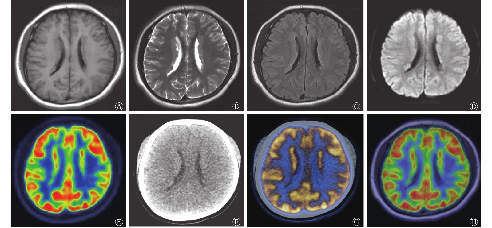
患者女性,16岁,因“间断突发晕厥一年余”于武汉大学人民医院就诊。既往史为初中体检发现贫血,具体不详,无其他特殊情况。实验室检查(大便常规、尿常规、血常规、凝血功能、肝肾功能及电解质检查)结果未见明显异常。行MRI及18F-FDG PET/CT检查,MRI图像显示,双侧脑室旁白质区可见异位的灰质,呈稍长T1信号及等T2信号(图1A、B),T2水抑制反转恢复(FLAIR)图像显示略高信号,边缘模糊,部分病灶悬于室管膜,突向侧脑室,形成锯齿状边缘(图1C);扩散加权成像结果显示,双侧脑实质未见异常扩散受限病灶,相应表观扩散系数未减低(图1D)。18F-FDG PET/CT图像显示,沿双侧脑室旁见条形稍高密度影,放射性分布呈条形浓聚,密度及放射性摄取与大脑皮质相仿(图1E~G)。结合MRI与18F-FDG PET/CT图像,显示邻近侧脑室稍受压,双侧大脑及小脑的其他部位未见异常放射性浓聚影,皮层下各神经核团显影清晰,临床诊断为HGM(图1H)。予以抗癫痫药物治疗后,患者病情稳定。

图 1 大脑灰质异位患者(女性,16岁)的18F-FDG PET/CT-MRI三模式显像图
Figure 1. 18F-FDG PET/CT-MRI three-mode images of the patient with heterotopic gray matter (female, 16 years old)
-
MRI对灰白质具有高度分辨力,是诊断HGM最为灵敏的方法。本病例的MRI图像中双侧脑室旁白质区可见异位的灰质。根据MRI图像显示的病灶部位及其影像学特征,许多研究者提议将HGM分为室管膜下或侧脑室旁的结节型、多位于白质内呈片状改变的极型以及位于侧脑室和皮质间的带型,其中,室管膜下HGM较为常见;根据病灶的累及范围又可将HGM分为双侧弥漫性、双侧局限性和单侧局限性[1, 5]。此外,还有研究结果显示,根据MRI的影像学特征,HGM可分为小灶型(呈“小岛状”)、大灶型(呈“团块状”)和带状型(呈“带状影”),本病例属于带状型,且在所有MRI序列上皆表现为病灶与脑灰质信号相仿,无明显强化信号[6]。HGM需与淋巴瘤、转移瘤、沿室管膜生长的室管膜瘤或颅内肿瘤以及结节性硬化相鉴别,HGM与脑肿瘤的MRI鉴别要点在于后者呈T1低信号、T2高信号改变,若合并出血则T1、T2均呈高信号改变,且脑肿瘤会出现占位效应和病灶周围明显水肿,增强后病灶亦有不同程度特征的强化,而HGM无上述征象[6-7]。
MRI具有较高的软组织分辨率,可以显示病灶的部位、累及范围、形态以及与周围组织结构的关系,而PET/CT可以从功能代谢方面获得病灶的分子生物学特征等更多信息,且与其他恶性肿瘤相鉴别。HGM的PET/CT一般征象为病灶的放射性摄取与正常脑皮质相仿或略低,这可能是由异位的神经元未成熟,且数目较少、排列不齐以及胶质纤维增加所致。本病例所应用的PET/CT-MRI三模式显像技术可依靠MRI提供解剖学信息,18F-FDG PET/CT提供代谢信息,两者相互印证,互为补充,从而实现准确诊断,精准治疗。有研究结果显示,1例难治性部分复杂性癫痫发作的患者通过行PET/CT-MRI三模式显像诊断为HGM,其MRI图像显示右侧脑室枕角边缘异位灰质信号,18F-FDG PET/CT图像显示病灶放射性摄取增加[8]。本病例的18F-FDG PET/CT影像学特征与上述病例相仿,即沿双侧脑室旁见条形稍高密度影,放射性分布呈条形浓聚,密度及放射性摄取与大脑皮质相仿,但本病例的PET/CT-MRI融合图可以更加清晰地显示病灶邻近侧脑室稍受压。由此可见,PET/CT-MRI三模式显像结合了多种影像学检查方法的优势,既可以精准发现病灶的位置,又可以测定其累及部位代谢的变化,从而为后期治疗方案的制定提供可靠的影像学信息。
然而,在临床上,患者因诸多限制难以同时进行PET/CT和MRI扫描。此外,虽然在后期图像融合时软件可自动识别和集中匹配,或在融合视图中同时显示PET/CT和MRI数据集,并自动识别和调整融合图像,但是如果临床医师对配准结果不满意,则需手动平移或调整图像,从而引入一定误差,故此方法具有一定的局限性。近年来,PET/MRI一体机技术飞速发展,其具有更高的诊断效率和便利性,但检查费用昂贵、检查时间长及易产生伪影等问题限制了其在临床上的广泛使用[9]。随着PET/MRI技术的进一步发展,这些问题可能在不久的将来得到解决,总体而言,多模态显像技术具有广阔的临床应用前景,需要进一步探索与研究。
利益冲突 本研究由署名作者按以下贡献声明独立开展,不涉及任何利益冲突。
作者贡献声明 李雪蓉负责论文的撰写;李蒙负责图像的采集;涂宁、冯洪燕负责图像的后处理;卜丽红负责论文的审阅。
18F-FDG PET/CT-MRI三模式显像在大脑灰质异位诊断中的应用
Application of 18F-FDG PET/CT-MRI three-mode imaging in heterotopic gray matter
-
摘要: 大脑灰质异位(HGM)是由于胚胎时期神经元移行中途受阻而在异常部位聚集所引起的一种少见的先天发育异常。笔者通过对HGM患者行18F-氟脱氧葡萄糖(FDG)PET/CT-MRI三模式显像分析其影像学特征,以期提高对该病影像学表现的认识,从而进行准确诊断和及时治疗。Abstract: Heterotopic gray matter (HGM) is a rare congenital developmental abnormality caused by the accumulation of neurons in abnormal parts due to the blockage of neuronal migration in the embryonic stage. The authors analyzed the imaging characteristics of a patient with HGM by18F-fluorodeoxyglucose (FDG) PET/CT-MRI three-mode imaging, in order to improve the understandingof the imaging manifestations of the disease, so as to facilitate accurate diagnosis and timely treatment.
-
Key words:
-
[1] 张葵, 李胜利, 文华轩, 等. 胎儿脑灰质异位的产前诊断及文献回顾[J]. 南方医科大学学报, 2015, 35(12): 1770−1774. DOI: 10.3969/j.issn.1673-4254.2015.12.21.
Zhang K, Li SL, Wen HX, et al. Prenatal diagnosis of fetal gray matter heteropia in one case and literature review[J]. J South Med Univ, 2015, 35(12): 1770−1774. DOI: 10.3969/j.issn.1673-4254.2015.12.21.[2] 徐忠平, 谢惠芳, 刘振华. 脑灰质异位症6例报告[J]. 第一军医大学学报, 2001, 21(4): 278. DOI: 10.3321/j.issn:1673-4254.2001.04.033.
Xu ZP, Xie HF, Liu ZH. Report of 6 cases of gray matter heterotopia[J]. J South Med Univ, 2001, 21(4): 278. DOI: 10.3321/j.issn:1673-4254.2001.04.033.[3] Oegema R, Barkovich AJ, Mancini GMS, et al. Subcortical heterotopic gray matter brain malformations: classification study of 107 individuals[J]. Neurology, 2019, 93(14): e1360−e1373. DOI: 10.1212/WNL.0000000000008200. [4] Lohmror A, Choudhary R. Movement disorder and epilepsy in subependymal nodular heterotopia[J]. J Med Sci, 2017, 37(4): 172−174. DOI: 10.4103/jmedsci.jmedsci_5_17. [5] Cossu M, Mirandola L, Tassi L. RF-ablation in periventricular heterotopia-related epilepsy[J]. Epilepsy Res, 2018, 142: 121−125. DOI: 10.1016/j.eplepsyres.2017.07.001. [6] 赵凯宇, 李晓红. 大脑灰质异位症的临床表现及影像学特征[J]. 实用医学杂志, 2009, 25(12): 2031−2032. DOI: 10.3969/j.issn.1006-5725.2009.12.048.
Zhao KY, Li XH. Clinical manifestations and imaging features of cerebral gray matter heterotopia[J]. J Pract Med, 2009, 25(12): 2031−2032. DOI: 10.3969/j.issn.1006-5725.2009.12.048.[7] 李书家, 胡喜斌, 高克克, 等. PET及MRI诊断脑灰质异位二例[J]. 放射学实践, 2003, 18(9): 694. DOI: 10.3969/j.issn.1000-0313.2003.09.037.
Li SJ, Hu XB, Gao KK, et al. Two cases of diagnosis of heterotopic brain gray matter by PET and MRI[J]. Radiol Pract, 2003, 18(9): 694. DOI: 10.3969/j.issn.1000-0313.2003.09.037.[8] Modarresifar H, Ho L. Brain heterotopia[J]. Clin Nucl Med, 2009, 34(3): 151−152. DOI: 10.1097/RLU.0b013e3181966dd4. [9] Ehman EC, Johnson GB, Villanueva-Meyer JE, et al. PET/MRI: where might it replace PET/CT?[J]. J Magn Reson Imaging, 2017, 46(5): 1247−1262. DOI: 10.1002/jmri.25711. -

 点击查看大图
图(1)
计量
- 文章访问数: 3379
- HTML全文浏览量: 1943
- PDF下载量: 12