


-
核素敷贴治疗作为核医学科的常用治疗手段,对皮肤血管瘤、瘢痕增生、寻常疣等浅表性病变具有治疗彻底、不留印记、方法简单、经济、无痛无痒等特点,因而受到广大患者的欢迎[1-3]。90Sr-90Y敷贴器是目前使用较为广泛的核素治疗源, 90Sr核素衰变释放出0.55 MeV的β粒子后转变为90Y,90Y衰变成90Zr时又释放出2.28 MeV的β粒子,整个敷贴器平均能量为0.98 MeV,β粒子最大射程12.90 mm,射线综合作用深度2~3 mm[4-6]。在实际临床治疗中,由于缺乏专业设备及人员,基层医院普遍采用简易公式对90Sr-90Y敷贴治疗的时间进行估算,导致部分患者受照剂量不足或者超量,严重影响了疗效,因而急需一种快捷且方便的剂量验证手段。EBT3胶片作为全新一代的辐射自显影胶片,具有免冲洗、可裁剪、剂量范围宽和组织等效性好等优点,目前被广泛应用于临床放射治疗剂量的验证[7]。后续有研究者发现,EBT3胶片的光谱响应与兆伏级能量束流的剂量率及性质无相关性[8-10],在射线能量0.003 MeV至18 MeV范围内,其能量依赖性小于3%[11],这些特性使得EBT3胶片在光子线和电子线混合野的剂量采集中也被广泛应用[12]。
本研究通过直线加速器产生的光子线对EBT3胶片进行标准剂量照射,应用胶片扫描仪对辐照后的胶片进行透射扫描,生成灰度-剂量标准曲线。再使用敷贴器对EBT3胶片组进行定时照射,扫描测得灰度值,根据标准曲线转换即可获取实际辐照剂量和空间剂量分布。本研究以期在原始数据缺失和基层专业设备及人员缺乏的情况下直接测量出不同治疗距离的敷贴器照射源对应的剂量大小及等剂量线范围。
-
90Sr-90Y敷贴器(北京原子高科股份有限公司,直径6 mm):标准圆形横截面,治疗面平整光滑,实测活度1.41×10⁸ Bq(3.8 mCi),杂散γ、X射线比例<<1%。EBT3胶片(美国Ashland公司Gafchromic EBT3型):中央层0.03 mm、两侧0.125 mm、单片厚度0.28 mm、能量范围keV至MeV、均匀性≤±3%、分辨率0.005 mm,允许剂量范围1~4000 cGy,按照顺序裁剪成6 cm×6 cm的正方形20片、10 cm×10 cm的正方形18片并标记。
-
应用直线加速器(美国Varian公司Trilogy型),选择6 MV光子线能量,加速器机架角度0°、准直器角度0°、辐射光野5 cm×5 cm,射野平坦度和对称性均<3%。在等中心(源与测量点垂直距离为100 cm)条件下对EBT3胶片进行曝光处理,每间隔50~100 MU(Monitor Unit跳数,在最大剂量点1 MU=1 cGy)曝光一片。应用德国IBA公司 Dose1型剂量仪进行监测,辐射剂量0~500 cGy;应用胶片扫描仪(美国Vidar公司 DosimetryPro Advantage型),选用红光通道对每片胶片进行透射扫描;应用仪器自带Film Analyzer分析软件绘制辐射剂量和扫描灰度对应的标准曲线。
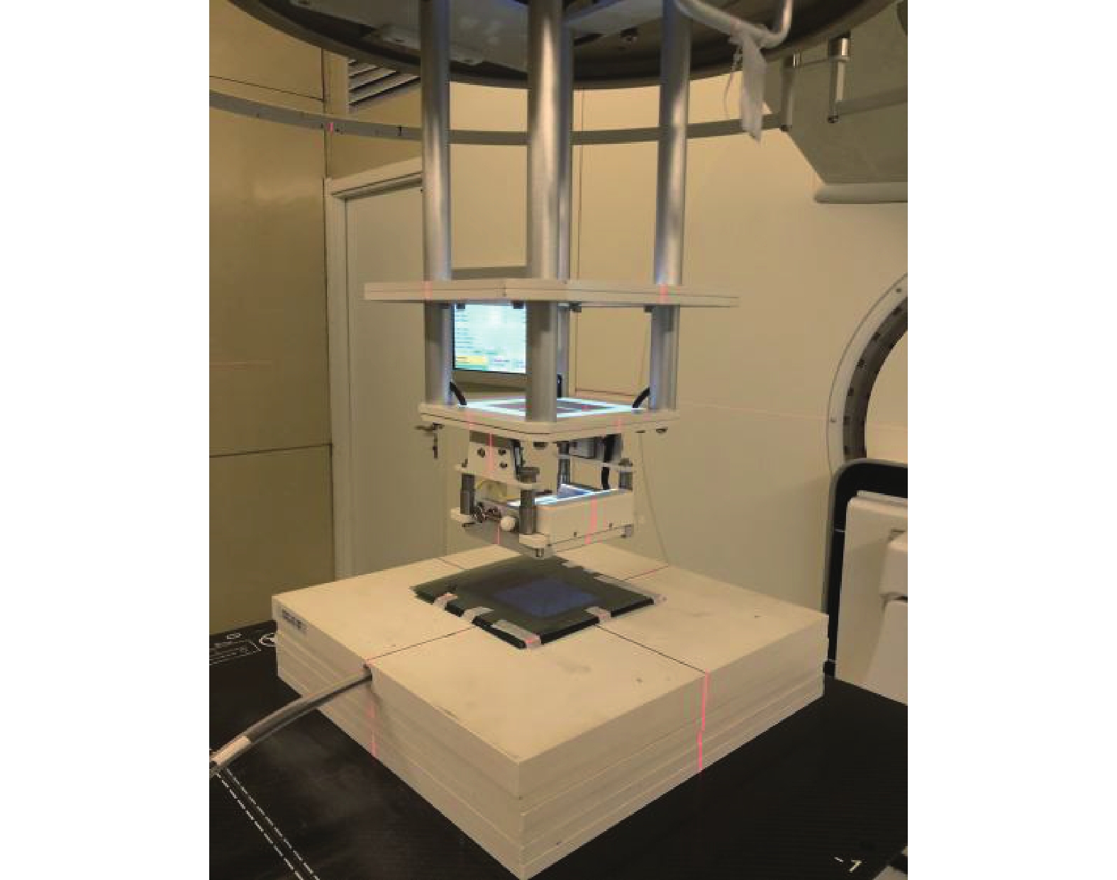
应用直线加速器按6、8、10、12、15 MeV能量分别出束,限光筒和标准挡块选择6 cm×6 cm,在标准源皮距(照射源与表面垂直距离100 cm)下使用固体水模(德国IBA公司Solid Phantom SP34型)分别在标准深度1.5 cm(6、8 MeV)和2.0 cm(10、12、15 MeV)处出束100 MU,应用Dose1剂量仪进行测量。将EBT3胶片叠加为厚度0.5 cm的胶片组(10 cm×10 cm,18片),分别替换原标准深度1.5 cm和2.0 cm处固体水模上的0.5 cm厚固体水模,在同等情况下,应用直线加速器各出束100 MU,记录测量数值(图1)。
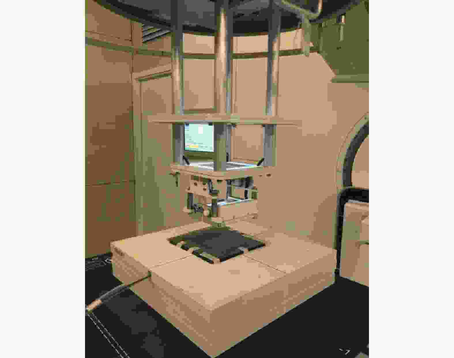
图 1 EBT3胶片的组织等效性测量
Figure 1. Measurement of EBT3 film tissue equivalence
-
将20片6 cm×6 cm的EBT3胶片重叠放置,使用游标卡尺测量,总厚度为5.6 mm,并用塑料夹具将其4个角固定。将90Sr-90Y敷贴器放置于胶片组顶层中央,照射4 min。胶片组经扫描后应用仪器自带Film Analyzer软件经过灰度-剂量标准曲线转换算出每张胶片中心轴附近的平均吸收剂量,根据胶片厚度推算出距离敷贴器照射源不同距离的中心轴辐射剂量,拟合剂量-深度跌落曲线。获取每一片胶片过中心轴所生成的一维剂量线,分别测量出50、100、150、200、250、300 cGy所覆盖区域的直径,应用仪器自带Matlab软件画出胶片的二维等剂量线示意图。
-
应用SPSS 20.0软件对数据进行统计学分析。符合正态分布的数据以
$\bar x \pm s $ 表示,在方差不齐的情况下,2种模体不同能量的电子线照射前后测得的吸收剂量的比较采用非参数独立样本U检验。P<0.05为差异有统计学意义。 -
加速器出束能量为6 MeV时,纯固体水模测量结果高于换入0.5 cm厚胶片后的测量结果,偏差为−2.17%;出束能量为8、10、12、15 MeV时,换入胶片后的测量结果均高于固体水模,偏差分别为1.24%、0.50%、0.68%和0.28%(表1)。由于6 MeV射线最大剂量深度为1.3 cm,测量深度为1.5 cm,导致剂量-深度轮廓线(profile)前移,故而实测剂量低于纯固体水模所测得剂量。其余能量实测深度均在最大深度点之前,0.5 cm厚胶片替换等厚度固体水模后测量结果略高于纯固体水模,但差异无统计学意义(Z=−0.31,P=0.84)。因此,利用胶片可以较为有效地模拟人体组织密度。
模体 6 MeV 8 MeV 10 MeV 12 MeV 15 MeV $ \bar x \pm s$ 

Z值 P值 固体水模(cGy) 95.93 99.42 100.90 103.60 106.80 101.33±4.13 −0.31 0.84 固体水模+EBT3胶片(cGy) 93.85 100.65 101.40 104.30 107.10 101.46±4.96 偏差(%) −2.17 1.24 0.50 0.68 0.28 − − − 注:EBT3胶片为10 cm×10 cm叠加18片的胶片组,厚度为0.5 cm。−表示无此项数据 表 1 不同能量照射后2种模体接受到的剂量比较
Table 1. Comparison of the doses received by the two phantoms irradiated after different energy
-
经过90Sr-90Y敷贴器照射4 min后的胶片组扫描图见图2,从1号胶片至20号胶片,随着距离的增加,辐射显影灰度随之降低,到20号时几乎与本底一致。

图 2 90Sr-90Y敷贴器照射4 min后重叠放置的20片EBT3胶片(6 cm×6 cm)每片的扫描图
Figure 2. Scanning images of 20 EBT3 films (6 cm×6 cm) placed overlappingly after 90Sr-90Y applicator irradiated for 4 min
-
由胶片扫描图2可见,胶片灰度在同一层面中心区域较为均匀,通过Film Analyzer分析软件进行灰度-剂量转换,测出不同深度处胶片对应的中心区域的平均剂量结果见表2。随着灰度的降低,剂量迅速下降,在0.5 cm深度处,剂量仅为7.0 cGy。
深度(mm) 剂量(cGy) 深度(mm) 剂量(cGy) 0 670.0 2.80 94.0 0.28 587.0 3.08 70.0 0.56 524.0 3.36 52.0 0.84 439.0 3.64 45.0 1.12 338.0 3.92 35.0 1.40 291.0 4.20 24.0 1.68 258.0 4.48 20.0 1.96 194.0 4.76 15.0 2.24 155.0 5.04 7.0 2.52 136.0 5.32 6.6 表 2 90Sr-90Y敷贴器照射后不同距离深度的剂量结果
Table 2. Dose results at different exposure distances after 90Sr-90Y applicator irradiation
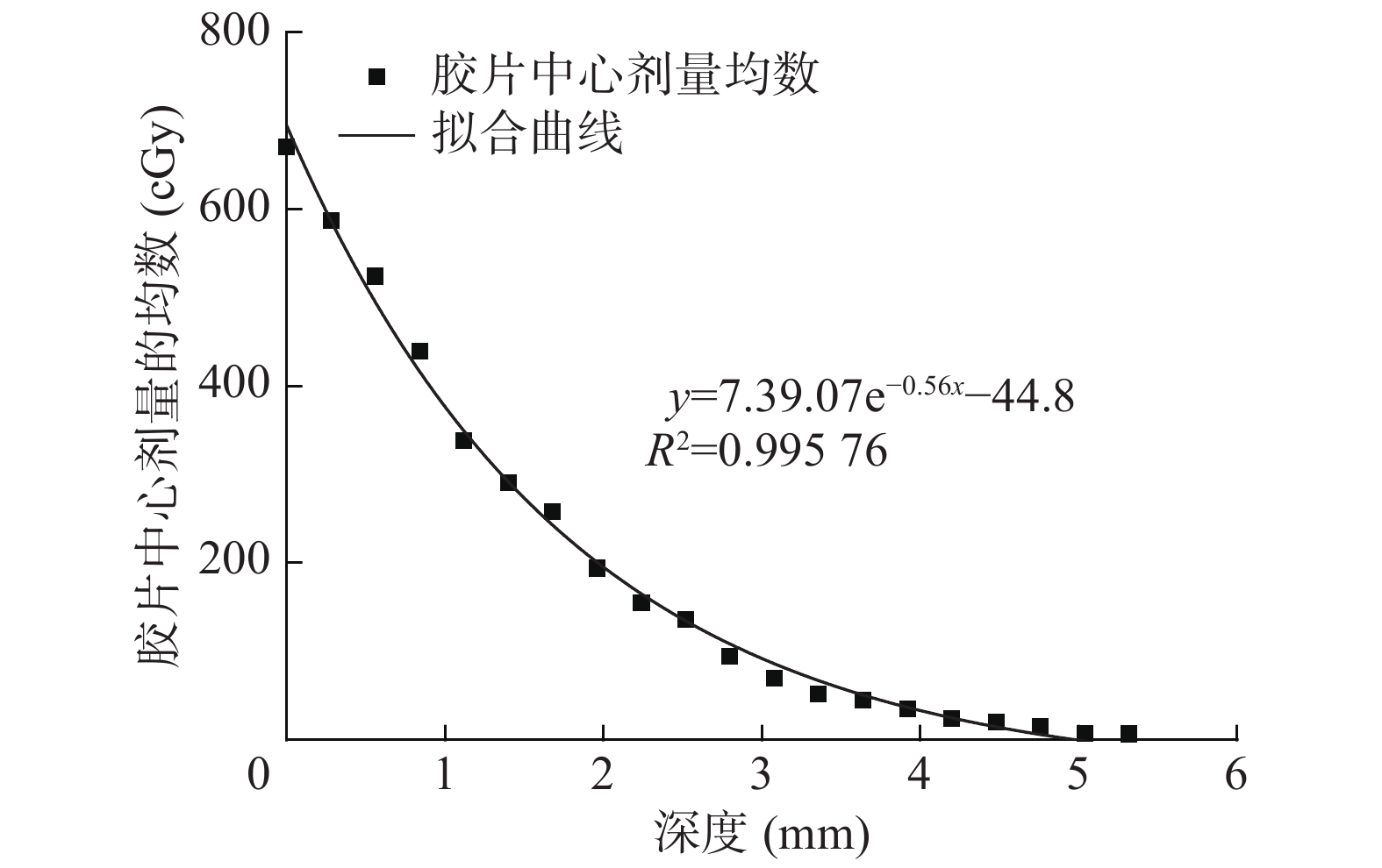
根据表2,拟合函数y=739.07e−0.56x−44.8,其中y表示距离敷贴器某一深度处中心轴的吸收剂量,单位cGy;x表示胶片距离敷贴器底面的垂直距离,单位mm。由图3可见,随着距离敷贴器深度的增加,辐射剂量呈指数下降的趋势,曲线拟合相关参数R2=0.99576。

图 3 90Sr-90Y敷贴器在EBT3胶片组内的照射深度与剂量拟合曲线
Figure 3. Fitting curve of irradiation depth and dose for 90Sr-90Y applicator in the EBT3 film group
-
不同剂量所覆盖胶片区域的直径结果见表3。胶片的二维等剂量线示意图见图4(俯视图)。可以看出1号胶片等剂量线间隔明显,随着等剂量线的降低,相互间的间距逐渐增加,而由过中心轴的等剂量线示意图4(剖面图)可以看出同一剂量所覆盖区域的直径大小随着距离敷贴器深度的增加均呈现先增大后减小的趋势。
胶片层数
(深度,mm)50 cGy 100 cGy 150 cGy 200 cGy 250 cGy 300 cGy 1(0) 10.3 9.2 8.5 8.2 8.0 7.8 2(0.28) 10.4 9.4 8.8 8.3 8.0 7.8 3(0.56) 10.5 9.5 8.9 8.4 8.0 7.6 4(0.84) 10.6 9.5 8.8 8.0 7.5 7.0 5(1.12) 10.6 9.6 8.5 7.8 6.7 6.3 6(1.40) 10.5 9.1 8.0 7.6 5.5 0 7(1.68) 10.5 9.0 7.7 5.7 3.9 0 8(1.96) 10.1 8.3 6.5 1.5 0 0 9(2.24) 9.5 7.2 4.0 0 0 0 10(2.52) 9.3 6.5 0 0 0 0 11(2.80) 7.6 1.2 0 0 0 0 12(3.08) 6.2 0 0 0 0 0 13(3.36) 3.2 0 0 0 0 0 14(3.64) 0 0 0 0 0 0 15(3.92) 0 0 0 0 0 0 表 3 90Sr-90Y敷贴器不同剂量不同胶片层数测量的区域直 径(mm)
Table 3. The area diameter measured by the 90Sr-90Y applicator at different doses and different number of film layers (mm)
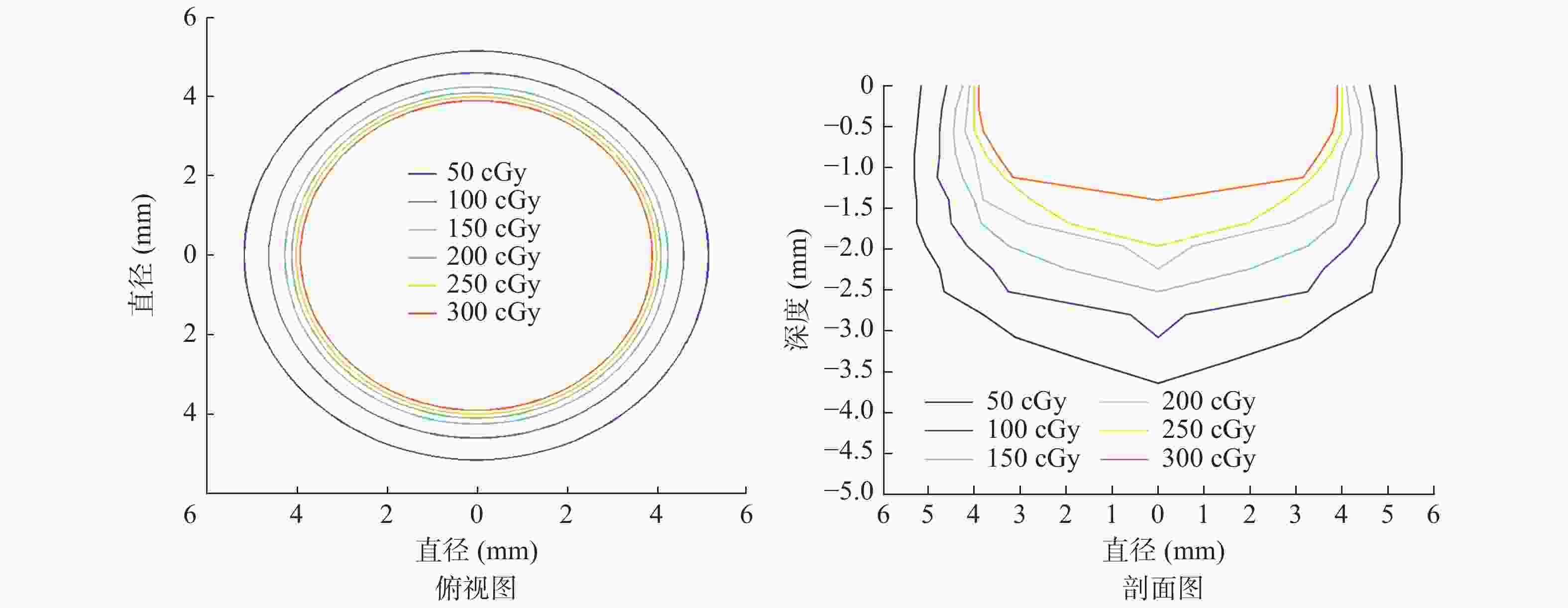
图 4 胶片平面二维等剂量线示意图
Figure 4. Schematic diagram of two-dimensional isodose lines in the film plane
-
90Sr-90Y敷贴器作为β射线面源(90Y核素退激产生的γ射线、内转换电子及韧致辐射产生的X射线占比<<1%,可以忽略不计[13]),平均能量为0.98 MeV,最大能量为2.28 MeV,属于连续能谱,能量分布范围较广,因而在实际临床应用时难以保证治疗剂量的准确性。目前临床普遍采用赵惠扬教授的计算方法(D0=0.93×1770×A/S)进行剂量率估算,取得了很好的临床效果[14],但公式中未完善敷贴器在组织不同深度处的空间等剂量分布。
Massillon-JI等[15]采用凝胶剂量测定法测定了90Sr-90Y粒子点源的三维剂量学特性,但没有测量90Sr-90Y面源的三维剂量特征。Hansen等[16]应用特制的小型外推电离室对眼科用90Sr-90Y敷贴源进行了有效的测量,但应用面较为局限,且测量设备需要特制。2009年施常备等[14]率先采用早期的辐射直接显色(RC)胶片做了32P面源持续低剂量率辐射敷贴治疗的胶片剂量验证,发现辐射直接显色胶片能够为32P敷贴源的剂量验证提供一定的帮助,但是目前关于90Sr-90Y源的胶片剂量验证研究较少。随着技术的进步,辐射直接显色胶片的能量响应、射线性质响应及剂量率响应等都有了极大的提高,因此本研究应用新型的EBT3胶片获取的90Sr-90Y面源的剂量分布,可为临床实践提供一定的参考。
本研究结果显示,应用EBT3胶片进行叠加剂量验证能够较准确地模拟人体组织。在剂量方面:敷贴器中心轴剂量跌落随距离敷贴器深度满足公式y=739.07e−0.56x−44.8,且呈指数衰减趋势,浅表处剂量最高,0.5 cm深度处中心轴剂量仅为表面剂量的1%,剂量曲线整体迅速跌落;敷贴器照射源中心轴剖面等剂量线分布随深度呈现先增大再迅速减小的趋势,常规4 min治疗200 cGy等剂量线所覆盖区域在浅表处为直径8.2 mm的圆面,而在治疗深度为1.96 mm时,其200 cGy等剂量线所覆盖区域直径不到2 mm,有效治疗面积仅为浅表处的1/16,难以覆盖治疗区域,分析原因是在浅表处受到敷贴器侧向散射及剂量建成区的双重影响,等剂量线展宽,而随着深度的增加,电子射程迅速降低,等剂量线随之迅速收窄。敷贴器照射源的这些特性无法通过经验公式得到,只能通过实际测量获取,而90Sr-90Y照射源,由于25 d就能达到衰变平衡,且整体半衰期为28.6年,1次测量就能够掌握敷贴器照射源较长时间范围内的剂量学特征,可有效地指导临床实践。
综上所述,对于临床应用的90Sr-90Y敷贴器,采用多层EBT3胶片测量能够获取其对应的剂量分布参数、拟合剂量-深度跌落曲线,获取对应深度的剂量线范围。根据所需治疗深度和范围,就可以较准确地计算治疗所需时间,这能为广大基层医疗机构核素敷贴剂量验证提供一种简单、快捷、较准确的方法。
利益冲突 本研究由署名作者按以下贡献声明独立开展,不涉及任何利益冲突。
作者贡献声明 杨涛负责方法的建立、现场的实验、论文的撰写;胡强负责数据的获取与分析;周云泷负责方法的建立、论文的审阅;金晓峰负责现场的实验、数据的获取;明慧负责文献的查阅、论文的审阅;余辉负责命题的提出、论文的审阅。
90Sr-90Y敷贴治疗的EBT3胶片剂量验证方法
EBT3 film dose verification method for 90Sr-90Y applicator
-
摘要:
目的 对90Sr-90Y敷贴治疗进行EBT3胶片剂量的测量,为临床应用提供一种快捷的剂量验证方法。 方法 选用能量响应较好的辐射直接显影EBT3胶片,并与固体水模进行比较,通过直线加速器建立0~500 cGy的胶片灰度-剂量标准曲线并检测其组织等效性。将20片6 cm×6 cm的EBT3胶片(每张胶片厚0.28 mm)重叠放置,将90Sr-90Y敷贴器放置在胶片最顶层,辐照4 min。通过灰度-剂量标准曲线计算出每张胶片中心轴附近的平均吸收剂量,根据胶片厚度计算距离敷贴器不同深度处的辐射剂量及等剂量线分布。采用非参数独立样本U检验比较不同能量的电子线照射前后所测得的吸收剂量的差异。 结果 胶片替换等厚度固体水模后测量结果略高于纯固体水模,但差异无统计学意义(Z=−0.31,P=0.84)。不同深度处胶片对应的中心区域随着灰度值的降低吸收剂量迅速下降,在0.5 cm深度处,剂量仅为7.0 cGy。4 min照射后,距离敷贴器不同深度处的吸收剂量满足拟合曲线y=739.07e−0.56x−44.8,等剂量线范围随深度的增加呈先增大后减小。 结论 EBT3胶片验证能够很好地满足90Sr-90Y敷贴治疗的剂量验证,具有简单、易行、准确的特点,适合基层医疗机构应用。 -
关键词:
- 胶片辐射剂量测定法 /
- 90Sr-90Y敷贴器 /
- EBT3胶片 /
- 剂量验证
Abstract:Objective To measure the EBT3 film dose of brachytherapy with 90Sr-90Y applicator to provide a rapid dose verification method for clinical application. Methods EBT3 films with good energy response were selected and compared with a solid water phantom. The gray scale dose curve of 0–500 cGy film was established using a linear accelerator, and its tissue equivalence was detected. Twenty 6 cm × 6 cm EBT3 films (0.28 mm thickness) were overlapped, and a 90Sr-90Y applicator was placed on the top and irradiated for 4 min. The absorbed dose near the central axis of each film was calculated using the gray scale dose curve. The radiation dose and isodose distributions at different distances from the applicator were calculated according to film thickness. Non-parametric independent sample U-test was used to compare the differences in absorbed doses measured before and after exposure to electron rays with different energies. Results The measurement results of film replacement with equal thickness solid water molds were slightly higher than those of pure solid water molds, but the difference was not statistically significant (Z=−0.31, P=0.84). The absorbed dose in the central region corresponding to different film depths decreased rapidly with decreasing gray values, and the dose at a depth of 0.5 cm was only 7.0 cGy. After irradiation for 4 min, the absorbed dose at different depths from the applicator met the fitting curve, y=739.07e−0.56x−44.8, and the range of isodose line initially increased and then decreased with the increasing depth. Conclusion EBT3 film verification can well meet the dose verification of 90Sr-90Y applicator, which is simple, easy, and accurate and is suitable of primary medical institutions. -
Key words:
- Film dosimetry /
- 90Sr-90Y applicator /
- EBT3 film /
- Dose verification
-
图 2 90Sr-90Y敷贴器照射4 min后重叠放置的20片EBT3胶片(6 cm×6 cm)每片的扫描图
Figure 2. Scanning images of 20 EBT3 films (6 cm×6 cm) placed overlappingly after 90Sr-90Y applicator irradiated for 4 min
图 3 90Sr-90Y敷贴器在EBT3胶片组内的照射深度与剂量拟合曲线
Figure 3. Fitting curve of irradiation depth and dose for 90Sr-90Y applicator in the EBT3 film group
图 4 胶片平面二维等剂量线示意图
Figure 4. Schematic diagram of two-dimensional isodose lines in the film plane
表 1 不同能量照射后2种模体接受到的剂量比较
Table 1. Comparison of the doses received by the two phantoms irradiated after different energy
模体 6 MeV 8 MeV 10 MeV 12 MeV 15 MeV $ \bar x \pm s$ Z值 P值 固体水模(cGy) 95.93 99.42 100.90 103.60 106.80 101.33±4.13 −0.31 0.84 固体水模+EBT3胶片(cGy) 93.85 100.65 101.40 104.30 107.10 101.46±4.96 偏差(%) −2.17 1.24 0.50 0.68 0.28 − − − 注:EBT3胶片为10 cm×10 cm叠加18片的胶片组,厚度为0.5 cm。−表示无此项数据  下载: 导出CSV
表 2 90Sr-90Y敷贴器照射后不同距离深度的剂量结果
Table 2. Dose results at different exposure distances after 90Sr-90Y applicator irradiation
深度(mm) 剂量(cGy) 深度(mm) 剂量(cGy) 0 670.0 2.80 94.0 0.28 587.0 3.08 70.0 0.56 524.0 3.36 52.0 0.84 439.0 3.64 45.0 1.12 338.0 3.92 35.0 1.40 291.0 4.20 24.0 1.68 258.0 4.48 20.0 1.96 194.0 4.76 15.0 2.24 155.0 5.04 7.0 2.52 136.0 5.32 6.6
下载: 导出CSV
表 3 90Sr-90Y敷贴器不同剂量不同胶片层数测量的区域直 径(mm)
Table 3. The area diameter measured by the 90Sr-90Y applicator at different doses and different number of film layers (mm)
胶片层数
(深度,mm)50 cGy 100 cGy 150 cGy 200 cGy 250 cGy 300 cGy 1(0) 10.3 9.2 8.5 8.2 8.0 7.8 2(0.28) 10.4 9.4 8.8 8.3 8.0 7.8 3(0.56) 10.5 9.5 8.9 8.4 8.0 7.6 4(0.84) 10.6 9.5 8.8 8.0 7.5 7.0 5(1.12) 10.6 9.6 8.5 7.8 6.7 6.3 6(1.40) 10.5 9.1 8.0 7.6 5.5 0 7(1.68) 10.5 9.0 7.7 5.7 3.9 0 8(1.96) 10.1 8.3 6.5 1.5 0 0 9(2.24) 9.5 7.2 4.0 0 0 0 10(2.52) 9.3 6.5 0 0 0 0 11(2.80) 7.6 1.2 0 0 0 0 12(3.08) 6.2 0 0 0 0 0 13(3.36) 3.2 0 0 0 0 0 14(3.64) 0 0 0 0 0 0 15(3.92) 0 0 0 0 0 0
下载: 导出CSV
-
[1] 赵洪伟, 阚侃, 吕茁, 等. 整形外科手术治疗瘢痕疙瘩的临床研究[J]. 中国卫生标准管理, 2020, 11(11): 43−45. DOI: 10.3969/j.issn.1674-9316.2020.11.016.
Zhao HW, Kan K, Lyu Z, et al. Clinical study of plastic surgery treatment of keloid tissue[J]. China Health Stand Manage, 2020, 11(11): 43−45. DOI: 10.3969/j.issn.1674-9316.2020.11.016.[2] 杨卷红, 张耀峰, 刘斌, 等. 核素32P简易敷贴治疗婴幼儿体表血管瘤的临床效果[J]. 临床医学研究与实践, 2020, 5(13): 109−110, 113. DOI: 10.19347/j.cnki.2096-1413.202013041.
Yang JH, Zhang YF, Liu B, et al. Clinical effect of nuclide 32P simple application in the treatment of infantile superficial hemangiomas[J]. Clin Res Pract, 2020, 5(13): 109−110, 113. DOI: 10.19347/j.cnki.2096-1413.202013041.[3] 潘展砚, 刘怡文. 32P敷贴治疗乳房外Paget病疗效评价[J]. 中国美容医学, 2017, 26(10): 77−78. DOI: 10.15909/j.cnki.cn61-1347/r.002002.
Pan ZY, Liu YW. The clinical evaluation of 32P-dressing radiotherapy in the treatment of extramammary Paget disease[J]. Chin J Aesthetic Med, 2017, 26(10): 77−78. DOI: 10.15909/j.cnki.cn61-1347/r.002002.[4] 黎凤明, 田晓东, 刘云瑛, 等. 联合整形外科手术及90Si-90Y放射性核素敷贴治疗瘢痕疙瘩的临床应用研究[J]. 中国临床医生杂志, 2016, 44(9): 32−34. DOI: 10.3969/j.issn.2095-8552.2016.09.012.
Li FM, Tian XD, Liu YY, et al. A clinical evaluation of plastic surgery combined with 90Si-90Y isotope stickers in the treatment of keloids[J]. Chin J Clin, 2016, 44(9): 32−34. DOI: 10.3969/j.issn.2095-8552.2016.09.012.[5] Rogers B, Lawrence J, Chmura J, et al. Dosimetric characterization of a novel 90Y source for use in the conformal superficial brachytherapy device[J]. Phys Med, 2020, 72: 52−59. DOI: 10.1016/j.ejmp.2020.03.002. [6] 郭晨雷, 张高龙, 钱建强. 利用磁谱仪测量90Sr-90Y放射源的β衰变能谱[J]. 物理与工程, 2011, 21(5): 10−12, 16. DOI: 10.3969/j.issn.1009-7104.2011.05.004.
Guo CL, Zhang GL, Qian JQ. Measurement of energy spectra for β decay of 90Sr-90Y by magnetic spectrometer[J]. Phys Eng, 2011, 21(5): 10−12, 16. DOI: 10.3969/j.issn.1009-7104.2011.05.004.[7] Hanušová T, Horáková I, Koniarová I. Pseudo-3D IMRT verification with EBT3 radiochromic film[J]. Radiat Prot Dosimetry, 2019, 186(2-3): 362−366. DOI: 10.1093/rpd/ncz232. [8] Sipilä P, Ojala J, Kaijaluoto S, et al. Gafchromic EBT3 film dosimetry in electron beams—energy dependence and improved film read-out[J/OL]. J Appl Clin Med Phys, 2016, 17(1): 360−373[2020-09-06]. https://aapm.onlinelibrary.wiley.com/doi/full/10.1120/jacmp.v17i1.5970. DOI: 10.1120/jacmp.v17i1.5970. [9] León-Marroquín EY, Mulrow DJ, Khan R, et al. Spectral analysis of the EBT3 radiochromic films for clinical photon and electron beams[J]. Med Phys, 2019, 46(2): 973−982. DOI: 10.1002/mp.13330. [10] Reinhardt S, Hillbrand M, Wilkens JJ, et al. Comparison of Gafchromic EBT2 and EBT3 films for clinical photon and proton beams[J]. Med Phys, 2012, 39(8): 5257−5262. DOI: 10.1118/1.4737890. [11] Hermida-López M, Lüdemann L, Flühs A, et al. Technical note: influence of the phantom material on the absorbed-dose energy dependence of the EBT3 radiochromic film for photons in the energy range 3 keV−18 MeV[J]. Med Phys, 2014, 41(11): 112103. DOI: 10.1118/1.4898598. [12] Sorriaux J, Kacperek A, Rossomme S, et al. Evaluation of Gafchromic® EBT3 films characteristics in therapy photon, electron and proton beams[J]. Phys Med, 2013, 29(6): 599−606. DOI: 10.1016/j.ejmp.2012.10.001. [13] 马俊杰, 韩寿岭, 张谦, 等. 锶-90/钇-90敷贴器γ、X射线的来源及所致医护人员的剂量[J]. 工业卫生与职业病, 2000, 26(3): 160−161. DOI: 10.3969/j.issn.1000-7164.2000.03.011.
Ma JJ, Han SL, Zhang Q, et al. Gamma-ray and X-ray from 90Sr/90Y applicator and its doses on medical staff[J]. Ind Health Occup, 2000, 26(3): 160−161. DOI: 10.3969/j.issn.1000-7164.2000.03.011.[14] 施常备, 梁晓燕, 袁彬, 等. 32P持续低剂量率辐射敷贴治疗的胶片剂量验证[J]. 现代肿瘤医学, 2009, 17(9): 1772−1774. DOI: 10.3969/j.issn.1672-4992.2009.09.061.
Shi CB, Liang XY, Yuan B, et al. The quantitative radiation dose by RC film for the continued low-dose-rate radiation of 32P[J]. J Mod Oncol, 2009, 17(9): 1772−1774. DOI: 10.3969/j.issn.1672-4992.2009.09.061.[15] Massillon-JL G, Minniti R, Mitch MG, et al. The use of gel dosimetry to measure the 3D dose distribution of a 90Sr/90Y intravascular brachytherapy seed[J]. Phys Med Biol, 2009, 54(6): 1661−1672. DOI: 10.1088/0031-9155/54/6/017. [16] Hansen JB, Culberson WS, DeWerd LA. Windowless extrapolation chamber measurement of surface dose rate from a 90Sr/90Y ophthalmic applicator[J]. Radiat Meas, 2018, 108: 34−40. DOI: 10.1016/j.radmeas.2017.11.0. -

 点击查看大图
计量
- 文章访问数: 4556
- HTML全文浏览量: 2834
- PDF下载量: 19
