


 下载:
下载:
-
肺肝样分化腺癌(hepatoid adenocarcinoma of the lung,HAL)是指肿瘤细胞具有肝细胞样分化、有类似肝细胞癌的形态特征、免疫组化甲胎蛋白(α-fetoprotein,AFP)表达呈阳性并且原发于肺部的腺癌[1]。HAL恶性程度高、侵袭性强、预后较普通型腺癌差,5年生存率约为55%[2]。此病临床罕见,多为个案报道,少有文献报道PET/CT显像的病例。我们对1例HAL患者的临床资料进行分析,结合文献总结其影像学特征,以提高医师对该病的认识。
-
患者男性,66岁,因“间断咳嗽伴痰中带血10个月,加重1周,少量咯血。”2018年9月于西安交通大学第一附属医院呼吸内科就诊。患者无发热、胸痛,无乏力、盗汗,有50年吸烟史,平均15支/d,有肿瘤家族遗传病史,父母分别因胃癌和淋巴瘤去世。实验室检查结果显示:AFP升高,为 28.49 ng/mL(0.00~7.00 ng/mL),癌胚抗原(CEA)、糖类抗原(carbohydrate antigen,CA)-125、CA-199、CA-724、总前列腺特异抗原(total prostate specific antigen,TPSA)均未见升高,结核杆菌定量测定为阴性。胸部CT平扫+增强扫描(图1)显示右肺上叶软组织肿块,未见明显分叶及毛刺征,增强CT扫描图像显示,病灶呈持续明显强化。18F-FDG PET/CT(图2)显示病灶的葡萄糖代谢水平显著升高,伴有局部坏死灶,考虑为恶性病变,全身未见转移征象。
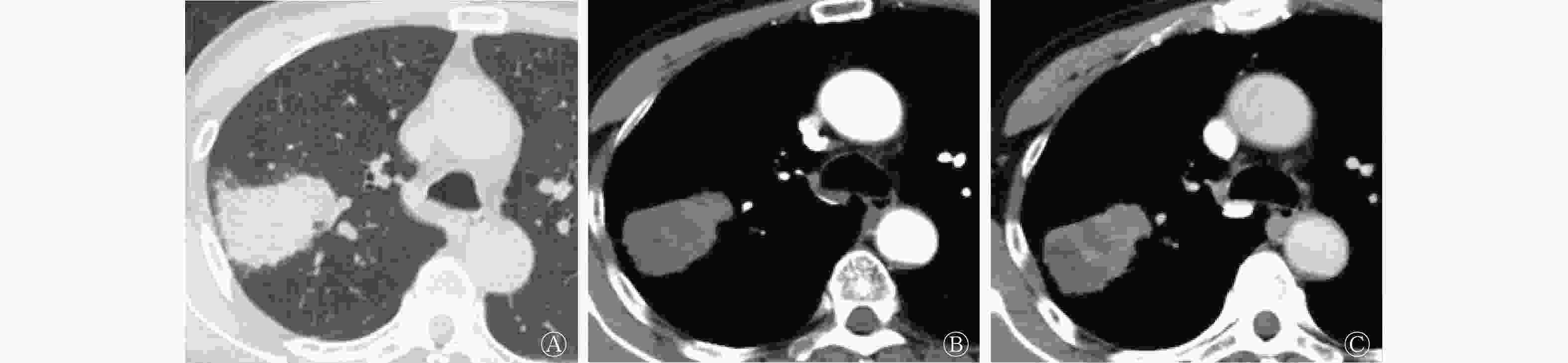
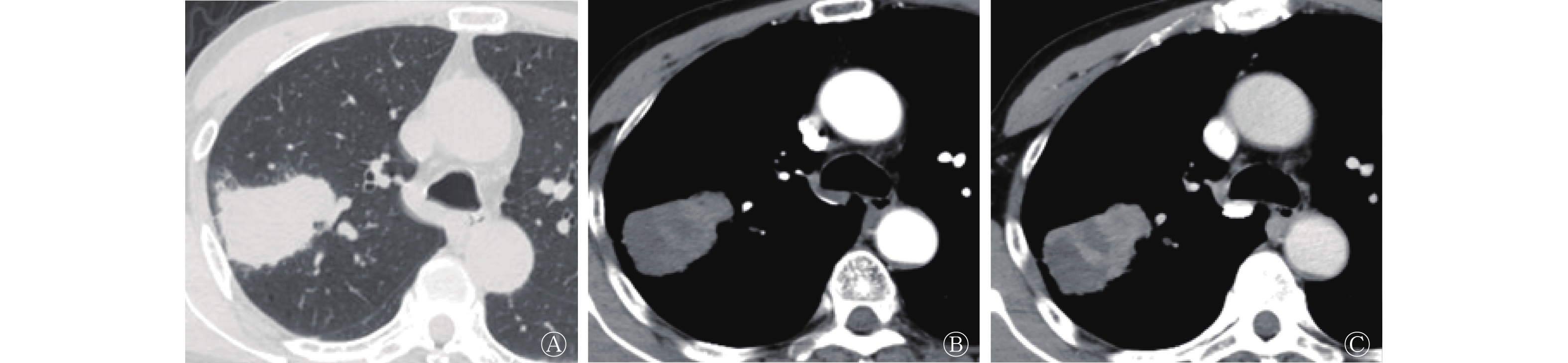
图 1 肺肝样分化腺癌患者(男性,66岁)的胸部CT平扫和增强扫描图 A、B、C分别为CT平扫、增强CT动脉期、增强CT静脉期图。CT平扫显示右肺上叶尖后段可见大小约4.1 cm×3.5 cm×4.0 cm的不规则软组织肿块影,边界清晰,内部密度不均匀,近肺门侧见斑片状稍高密度影;增强CT动脉期、静脉期病变明显且持续强化,远端片状低密度区未见明显强化。CT为计算机体层摄影术
Figure 1. Chest CT and contrast-enhanced CT images of a patient with hepatoid adenocarcinoma of the lung(male, 66 years old)
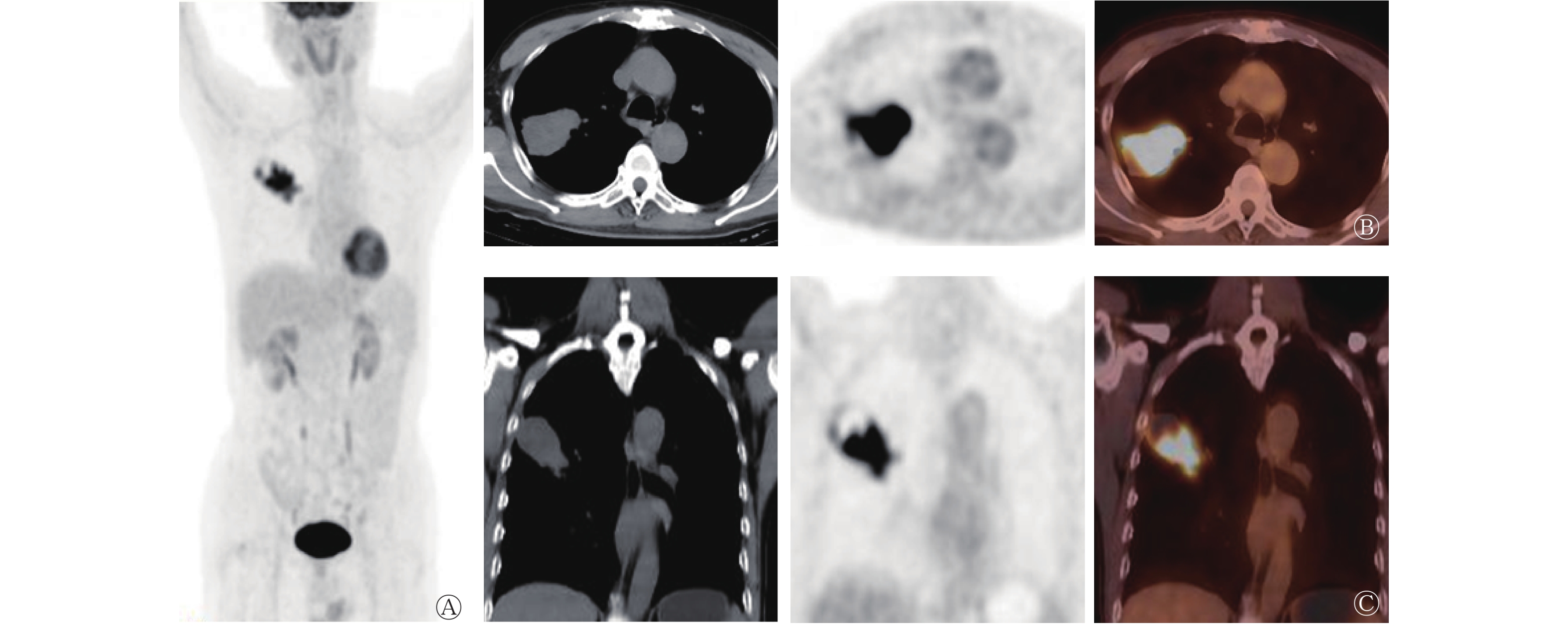
图 2 肺肝样分化腺癌患者(男性,66岁)的18F-FDG PET/CT显像图 A为最大密度投影图,示身体除病灶外的部位未见异常放射性核素浓聚灶;B、C分别为横断面和冠状面的CT、PET、PET/CT图,示右肺上叶不规则肿块,密度不均,实性部分靠近肺门侧,放射性核素摄取明显增高,SUVmax=14.2,远端低密度区放射性核素分布缺损。FDG为氟脱氧葡萄糖;PET为正电子发射断层显像术;CT为计算机体层摄影术;SUVmax为最大标准化摄取值
Figure 2. 18F-FDG PET/CT images of a patient with hepatoid adenocarcinoma of the lung(male, 66 years old)
入院后行右肺上叶肿块切除术,术后进行组织病理学检查发现,显微镜下见肿瘤组织呈巢团状排列,肿瘤细胞大且呈多边形、胞质丰富、免疫组化AFP呈阳性,Glypican-3、Hep Par1表达呈阳性,甲状腺转录因子1(thyroid transcription factor 1,TTF1)表达呈阴性(图3),符合HAL的特征,临床分期T2aN0M0。术后30 d后行培美曲塞+顺铂化疗,随访情况稳定。
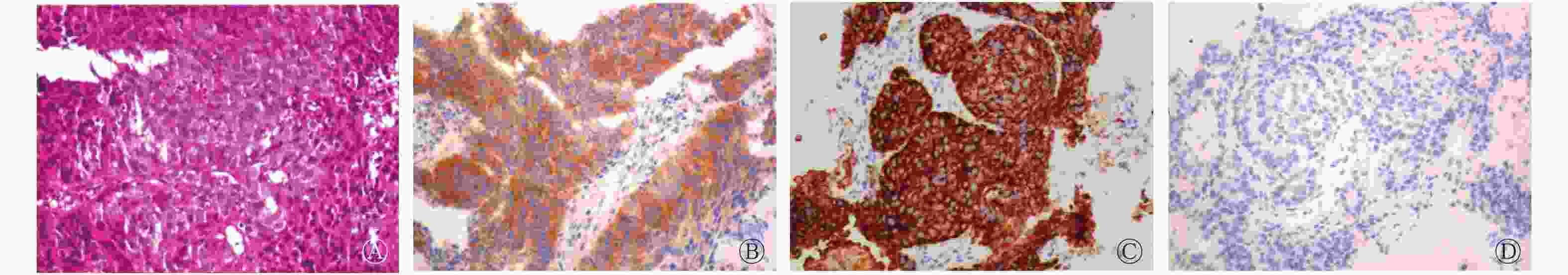
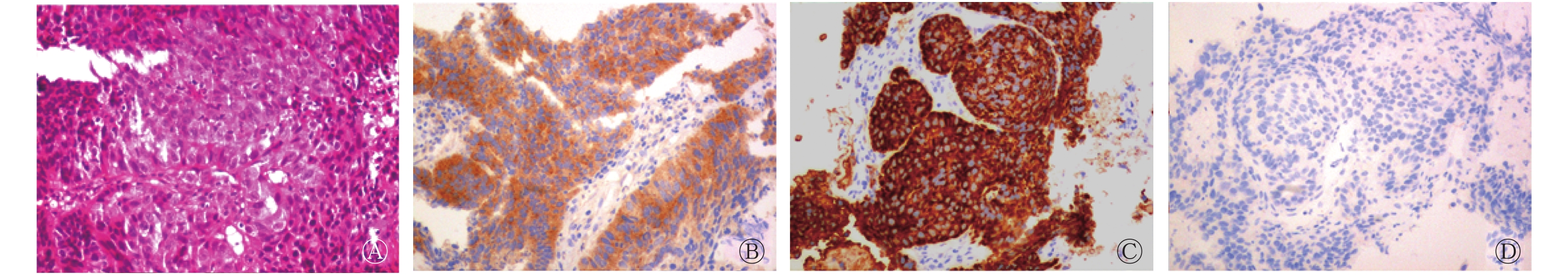
图 3 肺肝样分化腺癌患者(男性,66岁)的术后组织病理学检查(A)和免疫组织化学染色图(B~D) A为肺肝样分化腺癌患者的组织病理学检查图(苏木精-伊红染色,×200),显示癌细胞排列成团片状、异型显著;B~D分别为肺肝样分化腺癌患者的Glypican-3、Hep Par1、TTF1免疫组织化学染色图(×200),显示Glypican-3、Hep Par1表达呈阳性,TTF1表达呈阴性,符合肺肝样分化腺癌的特征。Glypican-3表达于胚胎性肝及胎盘组织的合体滋养叶细胞层中,一般用于鉴别肝细胞癌和肝脏良性增生结节。Hep Par1为肝癌细胞特异性标记;TTF1为甲状腺转录因子1
Figure 3. Postoperative pathological pictures and immunohistochemical staining of a patient with hepatoid adenocarcinoma of the lung(male, 66 years old)
-
HAL是一种具有腺癌及肝分化特征的肺癌类型,组织病理学检查与免疫组织化学检查是诊断HAL的必要条件[3]。血清AFP水平可作为诊断HAL的辅助条件,但也有文献报道AFP水平不升高[4-5]。丁前江等[6]报道了一例HAL中年男性患者,既往有吸烟史,体检结果显示右肺上叶有肿块,边界清晰,其内见坏死区,增强CT扫描图像明显强化。本例患者咳嗽伴咯血,咯血原因可能是肿块较大,导致内部出血。胸部CT平扫+增强扫描显示恶性病变,结合血清AFP水平升高,临床考虑为HAL,行18F-FDG PET/CT检查的目的是协助原始分期,确定下一步的治疗方案。18F-FDG PET/CT图像显示,病灶的葡萄糖代谢水平明显升高,我们预测疾病的恶性程度较高,未见远处转移,行肿瘤根治术后进行辅助化疗。
HAL的影像学表现以恶性征象为主,胸部CT平扫+增强扫描是诊断与鉴别肺部病变的首选方法,胸部CT平扫表现为较大的软组织肿块,边界欠清晰,密度不均匀,中心可见片状坏死区,增强CT扫描显示实性部分明显强化,坏死区不强化。18F-FDG PET/CT并非诊断HAL的必要方法,其主要表现为肿块实性部分葡萄糖代谢水平明显升高,病灶坏死部分葡萄糖代谢水平不增高或无明显增高,难以与其他类型的肿瘤、感染及肉芽肿病变相鉴别。18F-FDG PET/CT的独特优势体现在可以对恶性肿瘤进行原始分期、再分期、预后及早期疗效评价。本例患者通过全身18F-FDG PET/CT显像未发现远处转移,从而为临床行肺癌根治术提供了可靠依据。
利益冲突 本研究由署名作者按以下贡献声明独立开展,不涉及任何利益冲突。
作者贡献声明 沈聪负责资料的收集、论文的撰写;段小艺负责论文的修订。
肺肝样分化腺癌18F-FDG PET/CT显像一例
18F-FDG PET/CT imaging of hepatoid adenocarcinoma of the lung: a case report
-
摘要: 笔者报道了1例肺肝样分化腺癌(HAL)患者的18F-氟脱氧葡萄糖(FDG) PET/CT 显像,分别从临床症状、实验室检查、术前增强CT显像、手术所见及18F-FDG PET/CT显像等方面分析了该病的特点,并通过文献回顾总结了HAL的组织病理学及影像学特征。HAL在临床上较为罕见,多为个案报道,目前少有文献报道HAL18F-FDG PET/CT显像的病例。Abstract: The author reported a case of hepatoid adenocarcinoma of the lung (HAL) with 18F-fluorodeoxyglucose (FDG) PET/CT imaging, which was analyzed from clinical symptoms, laboratory examinations, preoperative contrast-enhanced CT imaging, surgical findings, and 18F-FDG PET/CT imaging. The pathological and imaging characteristics of HAL were summarized through literature review. Cases of HAL is clinically rare, and most of them are case reports. There are few case reports of 18F-FDG PET/CT imaging.
-
Key words:
-
图 1 肺肝样分化腺癌患者(男性,66岁)的胸部CT平扫和增强扫描图 A、B、C分别为CT平扫、增强CT动脉期、增强CT静脉期图。CT平扫显示右肺上叶尖后段可见大小约4.1 cm×3.5 cm×4.0 cm的不规则软组织肿块影,边界清晰,内部密度不均匀,近肺门侧见斑片状稍高密度影;增强CT动脉期、静脉期病变明显且持续强化,远端片状低密度区未见明显强化。CT为计算机体层摄影术
Figure 1. Chest CT and contrast-enhanced CT images of a patient with hepatoid adenocarcinoma of the lung(male, 66 years old)
图 2 肺肝样分化腺癌患者(男性,66岁)的18F-FDG PET/CT显像图 A为最大密度投影图,示身体除病灶外的部位未见异常放射性核素浓聚灶;B、C分别为横断面和冠状面的CT、PET、PET/CT图,示右肺上叶不规则肿块,密度不均,实性部分靠近肺门侧,放射性核素摄取明显增高,SUVmax=14.2,远端低密度区放射性核素分布缺损。FDG为氟脱氧葡萄糖;PET为正电子发射断层显像术;CT为计算机体层摄影术;SUVmax为最大标准化摄取值
Figure 2. 18F-FDG PET/CT images of a patient with hepatoid adenocarcinoma of the lung(male, 66 years old)
图 3 肺肝样分化腺癌患者(男性,66岁)的术后组织病理学检查(A)和免疫组织化学染色图(B~D) A为肺肝样分化腺癌患者的组织病理学检查图(苏木精-伊红染色,×200),显示癌细胞排列成团片状、异型显著;B~D分别为肺肝样分化腺癌患者的Glypican-3、Hep Par1、TTF1免疫组织化学染色图(×200),显示Glypican-3、Hep Par1表达呈阳性,TTF1表达呈阴性,符合肺肝样分化腺癌的特征。Glypican-3表达于胚胎性肝及胎盘组织的合体滋养叶细胞层中,一般用于鉴别肝细胞癌和肝脏良性增生结节。Hep Par1为肝癌细胞特异性标记;TTF1为甲状腺转录因子1
Figure 3. Postoperative pathological pictures and immunohistochemical staining of a patient with hepatoid adenocarcinoma of the lung(male, 66 years old)
-
[1] Zhuansun YX, Bian LJ, Zhao ZX, et al. Clinical characteristics of hepatoid adenocarcinoma of the lung: four case reports and literature review[J]. Cancer Treat Res Commun, 2021, 12(29): 100474. DOI: 10.1016/j.ctarc.2021.100474. [2] Tonyali O, Gonullu O, Ozturk MA, et al. Hepatoid adenocarcinoma of the lung and the review of the literature[J]. J Oncol Pharm Pract, 2020, 26(6): 1505−1510. DOI: 10.1177/1078155220903360. [3] Hou Z, Xie J, Zhang L, et al. Hepatoid adenocarcinoma of the lung: a systematic review of the literature from 1981 to 2020[J/OL]. Front Oncol, 2021, 11: 702216[2021-08-16]. https://pubmed.ncbi.nlm.nih.gov/34422656/. DOI: 10.3389/fonc.2021.702216. [4] Sun JN, Zhang BL, Li LK, et al. Hepatoid adenocarcinoma of the lung without production of α-fetoprotein: a case report and review of the literature[J]. Oncol Lett, 2016, 12(1): 189−194. DOI: 10.3892/ol.2016.4559. [5] Haninger DM, Kloecker GH, Bousamra Ii M, et al. Hepatoid adenocarcinoma of the lung: report of five cases and review of the literature[J]. Mod Pathol, 2014, 27(4): 535−542. DOI: 10.1038/modpathol.2013.170. [6] 丁前江, 邓茜, 李凯敏, 等. 肺肝样腺癌1例[J]. 中华胸心血管外科杂志, 2019, 35(11): 700−701. DOI: 10.3760/cma.j.issn.1001-4497.2019.11.014.
Ding QJ, Deng Q, Li KM, et al. Hepatoid adenocarcinoma of the lung: one case report[J]. Chin J Thorac Cardiovasc Surg, 2019, 35(11): 700−701. DOI: 10.3760/cma.j.issn.1001-4497.2019.11.014. -

 点击查看大图
计量
- 文章访问数: 3237
- HTML全文浏览量: 2428
- PDF下载量: 20