


 下载:
下载:
-
肋骨血管瘤是一种较罕见的骨肿瘤,通常与恶性骨肿瘤难以区分,其发病率不到恶性骨肿瘤的1%[1]。肋骨血管瘤常被误诊为原发性或继发性恶性骨肿瘤。由于60%~80%的原发性肋骨肿瘤均为恶性[1],因此了解肋骨血管瘤的影像学特征至关重要。我们分析了 1 例临床确诊为肋骨血管瘤患者的影像学表现特点及鉴别诊断过程,旨在为临床诊治提供参考。
-
患者女性,77岁,因“左侧背部疼痛10余天”为主诉就诊。患者于10 天前出现左侧背部疼痛,呈间断性刺痛,3天前就诊于当地医院,行胸部CT检查,结果显示:左侧第6后肋膨胀性骨质破坏,突破骨皮质,周围可见软组织肿块,其内可见多发高密度。行“抗炎”治疗后疼痛稍缓解,为求进一步诊治而就诊于河北医科大学第三医院骨科。患者一般情况良好,否认家族遗传病史。体格检查结果:胸廓无畸形,两侧对称,肋间隙无异常增宽或缩窄,两侧呼吸动度一致,触觉语颤均等,无增强或减弱,胸壁未扪及明显肿块,左后背部有轻触痛。两肺叩诊清音,肺肝相对浊音界位于右锁骨中线第5肋间,两肺呼吸音清,未闻及干、湿性啰音及胸膜摩擦音。实验室检查结果:血尿便常规、肝肾功能、电解质及肿瘤标志物未见明显异常。
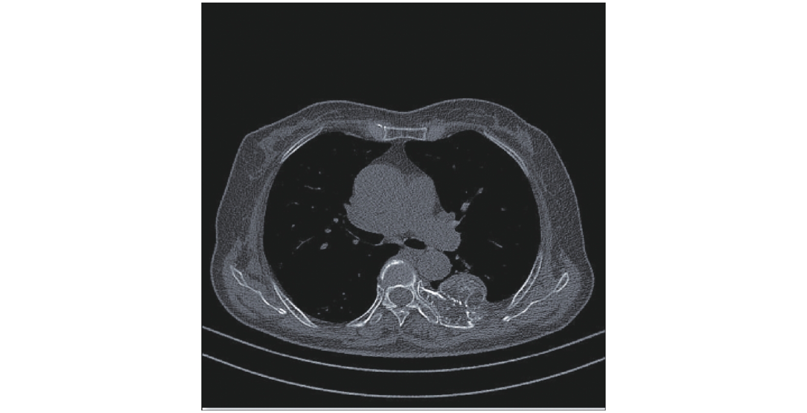
胸部CT及第5~7肋骨CT显像结果:左侧第6后肋骨膨胀性骨质破坏,其内密度不均匀,可见斑点状高密度影,后肋骨内侧可见骨性突起,局部与肋骨髓腔相通,内可见斑点状高密度影,局部皮质连续性欠佳,邻近左侧第5后肋骨局部骨质受压,周围软组织稍厚,邻近胸膜肥厚,考虑为肋骨恶性肿瘤(图1)。
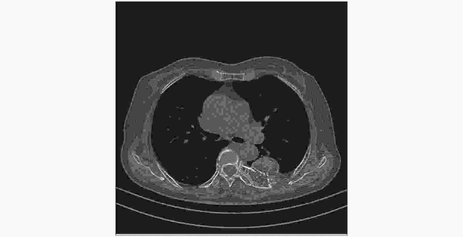
图 1 肋骨血管瘤患者(女性,77岁)的胸部CT图
Figure 1. Chest CT image of the patient with rib hemangioma (female, 77 years old)
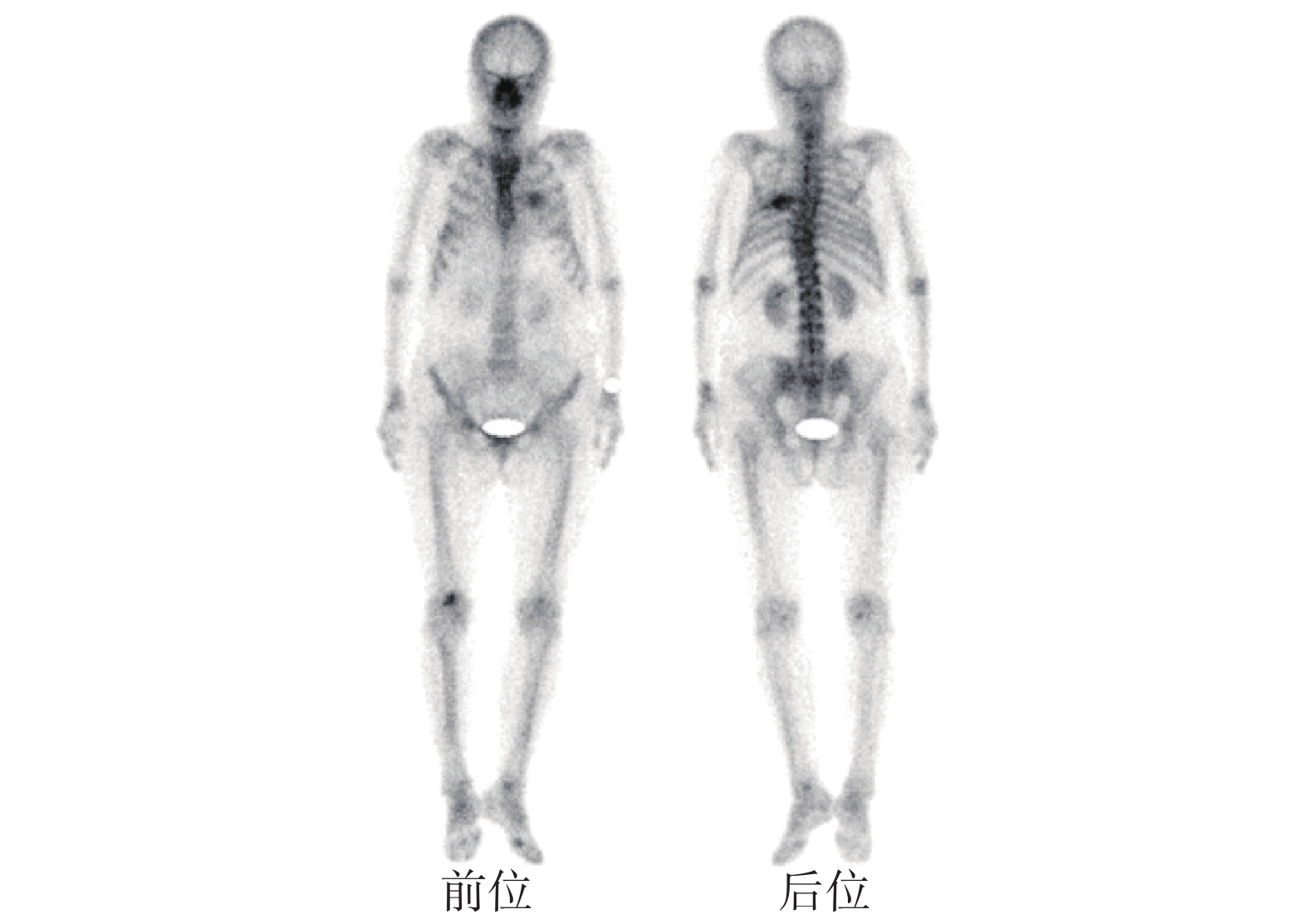
为协助诊断和评估病变范围,患者行99Tcm-MDP(MDP由北京师宏药物研究中心提供)SPECT(美国GE公司Intinia Hawkey型)全身骨显像,结果:左后第6肋骨形态欠规整且伴异常放射性浓聚,余骨未见明显异常放射性浓聚或稀疏区(图2)。

图 2 肋骨血管瘤患者(女性,77岁)的99Tcm-MDP SPECT全身骨显像图(前、后位)
Figure 2. 99Tcm-MDP SPECT whole body bone imaging of rib hemangioma (female, 77 years old)
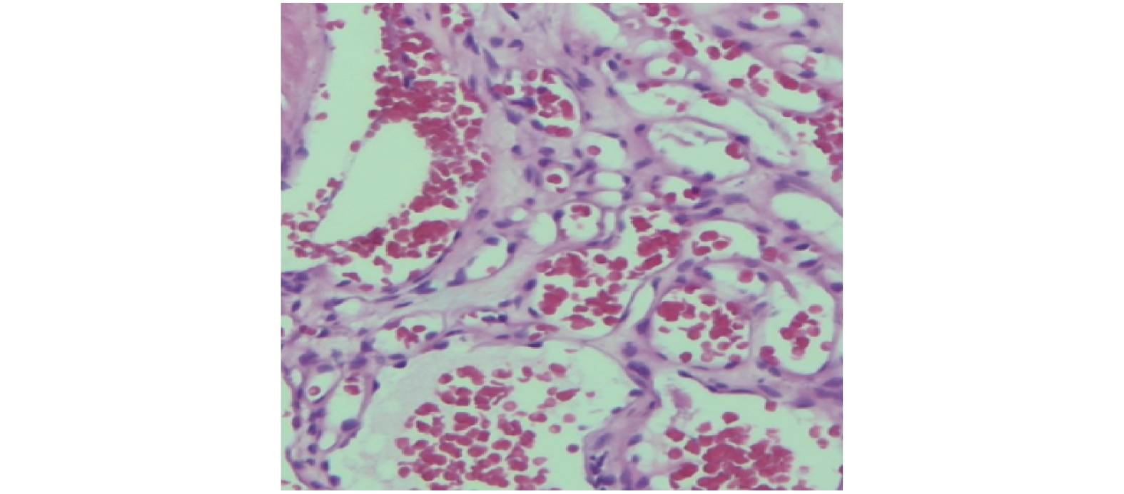
患者于全麻下行左侧肋骨肿物扩大切除术+胸壁缺损重建术+肋间神经阻滞术,术中所见:左侧第6后肋骨可见肋骨肿物,大小约4 cm×4 cm,外形不规则、质硬、边界清、包膜完整、血运丰富。术后组织病理学检查结果:骨小梁间脂肪组织中可见簇状分布的大小不等的团灶状血管,诊断为肋骨血管瘤(图3)。患者于术后半年内行CT随访,病灶愈合良好。

图 3 肋骨血管瘤患者(女性,77岁)的组织病理学检查图(苏木精-伊红染色,×200)
Figure 3. Histopathological examination of the patient with rib hemangioma(female, 77 years old)
-
肋骨肿瘤在临床中比较常见,其中以骨转移瘤多见,而肋骨良性肿瘤中,以骨软骨瘤居多,肋骨血管瘤极为罕见[1-3]。骨血管瘤为原发于骨骼血管的良性肿瘤,是一种呈瘤样增生的血管组织,掺杂于骨小梁之间,不易单独分离[1]。该病可发生于任何年龄,发病高峰在50~60岁,女性好发,大多数患者没有明显的临床症状,多为偶然发现,少部分患者可出现局限性胸背部疼痛等不适。骨血管瘤的好发部位是椎体和颅骨,约占骨血管瘤的2/3,其次为四肢长骨,其他部位少见,肋骨血管瘤则极为罕见,临床偶有报道[1-3]。本例患者为77岁女性,主要症状为左侧背部间断性刺痛,CT图像显示左侧第6后肋骨膨胀性骨质破坏,内见斑点状高密度影,考虑为恶性病变的可能性较大,不除外为骨转移瘤的可能;患者为了确诊是否为全身骨转移,遂行全身骨显像,结果显示左后第6肋骨形态欠规整且伴异常放射性浓聚,余处未见明显异常浓聚灶。我们结合患者的病史,首先考虑为肋骨单发骨肿瘤,然后经手术切除、术后组织病理学检查结果为肋骨血管瘤。
虽然肋骨血管瘤的最终确诊需要依靠组织病理学检查,但是影像学检查在疾病的诊断和鉴别诊断过程中依然能够发挥一定的作用。X射线及CT显像表现为受累肋骨呈梭形膨胀,多见于向胸内突出,可见边界清楚的骨质破坏区,其内可见稀疏而粗乱的骨纹理或呈放射状排列的骨小梁,恶变者骨皮质连续性中断,周围有软组织肿块。因此,CT三维重建有利于肿瘤的明确定位[4]。
全身骨显像可以通过一次检查而显示出全身骨骼的病变情况,其对于多骨病变,尤其是对恶性肿瘤骨转移的诊断具有较好的临床价值。同时,骨显像剂摄取程度的高低对显示肋骨病变的范围和鉴别良、恶性肿瘤等方面也有一定的参考价值。目前,SPECT/CT设备日益普及,其能够结合病灶摄取骨显像剂的变化和CT中病灶的解剖结构的变化,对于肋骨病变的诊断和鉴别诊断可发挥更加重要的作用。
肋骨血管瘤需与以下疾病相鉴别。(1)肋骨骨纤维结构不良:患者多发病于0~20岁,在X射线和CT显像中表现为皮质内单发或多发病变,多有硬化边,受累皮质膨胀变薄或增厚,病灶内部不均匀,可见点状钙化及类似磨玻璃样密度,无骨膜反应[5]。在骨显像中,典型的骨纤维结构不良为沿肋骨走行的条状异常放射性浓聚灶。(2)肋骨转移瘤:患者发病年龄一般偏大,多患有原发恶性肿瘤,常表现为多发成骨性或者破骨性骨质异常(多发肋骨病变与其他骨骼同时受累等),骨显像中可见全身骨骼无规律分布的多发异常放射性浓聚区或稀疏缺损区。(3)肋骨软骨肉瘤:患者多为20岁以上的成年人,多发生于肋与肋软骨结合部,病灶边界欠清,其内可见多发致密弧形及斑点状钙化,骨皮质连续性中断,周围可见少量软组织密度影,全身骨显像中病灶表现为单发的异常放射性浓聚灶。(4)骨巨细胞瘤:患者多发病于20~40岁,影像学表现为溶骨性骨破坏,病变边界较清,无硬化边,骨皮质变薄,局部中断,病变内密度欠均匀[6]。全身骨显像中病灶表现为团块状异常放射性浓聚灶。
综上所述,肋骨血管瘤的诊断应根据临床症状、影像学表现、临床诊断及组织病理学检查进行综合判断,SPECT对于该病的诊断和鉴别诊断具有一定的参考价值。
利益冲突 本研究由署名作者按以下贡献声明独立开展,不涉及任何利益冲突。
作者贡献声明 贾桐负责研究命题的提出、方法的建立、数据的分析、论文的撰写;黄建敏、魏玲格负责论文的审核与修订;傅鹏、解朋负责数据的采集、现场工作的实施。
肋骨血管瘤骨显像一例
A case report of rib hemangioma demonstrated by bone scintigraphy
-
摘要: 笔者报道了1例肋骨血管瘤患者,即从临床症状、影像学表现、临床诊断、组织病理学检查诊断及预后等方面介绍并分析了该病的特点,并通过文献复习加深了对肋骨血管瘤的认识。该病为肋骨的血管瘤病变,其进展相对较缓慢,虽然在影像学上无明显的特异性表现,但并非只能通过组织病理学检查诊断,影像学表现在该病的诊断上仍有一定的参考价值。该病的影像学表现多种多样,易与恶性骨肿瘤及骨肿瘤样病变混淆。因该病极易误诊,所以需结合临床表现、影像学检查及术后组织病理学检查综合考虑。Abstract: A case of rib hemangioma was reported. The characteristics of the disease were analyzed including the clinical symptoms, imaging manifestations, clinical diagnosis, pathological diagnosis and histopathological, and were deepened the understanding through literature review. The case was confirmed as rib hemangioma by the pathology. The disease progresses relatively slowly and had low specificity in the imaging. It was not only diagnosed by histopathological, but the imaging findings still have some value in this disease. The imaging manifestations of the disease are varied, which are easy to be confused with malignant bone tumors and bone tumor-like lesions. Once myofibroblastoma occurs, it is easy to misdiagnose. Thus, a combination of clinical symptoms, imaging manifestations, postoperative histopathological and therapeutic considerations is needed.
-
Key words:
-
图 1 肋骨血管瘤患者(女性,77岁)的胸部CT图
Figure 1. Chest CT image of the patient with rib hemangioma (female, 77 years old)
图 2 肋骨血管瘤患者(女性,77岁)的99Tcm-MDP SPECT全身骨显像图(前、后位)
Figure 2. 99Tcm-MDP SPECT whole body bone imaging of rib hemangioma (female, 77 years old)
-
[1] Haro A, Nagashima A. A rare case report of rib hemangioma mimicking a malignant bone tumor or metastatic tumor[J/OL]. Int J Surg Case Rep, 2015, 16: 141–145[2020-03-26]. https://www.ncbi.nlm.nih.gov/pmc/articles/PMC4643469. DOI: 10.1016/j.ijscr.2015.09.036. [2] Tew K, Constantine S, Lew WYC. Intraosseous hemangioma of the rib mimicking an aggressive chest wall tumor[J]. Diagn Interv Radiol, 2011, 17(2): 118−121. DOI: 10.4261/1305-3825.DIR.3031-09.2. [3] Okumura T, Asamura H, Kondo H, et al. Hemangioma of the rib: a case report[J]. Jpn J Clin Oncol, 2000, 30(8): 354−357. DOI: 10.1093/jjco/hyd097. [4] Yuan Y, Matsumoto T, Miura G. Imaging findings of an intercostal hemangioma[J]. J Thorac Imaging, 2002, 17(1): 92−95. DOI: 10.1097/00005382-200201000-00015. [5] Khanna M, Delaney D, Tirabosco R, et al. Osteofibrous dysplasia, osteofibrous dysplasia-like adamantinoma and adamantinoma: correlation of radiological imaging features with surgical histology and assessment of the use of radiologyi in contributing to needle biopsy diagnosis[J]. Skeletal Radiol, 2008, 37(12): 1077−1084. DOI: 10.1007/s00256-008-0553-1. [6] Hunter CL, Pacione D, Hornyak M, et al. Giant-cell tumors of the cervical spine: case report[J]. Neurosurgery, 2006, 59(5): E1142−E1143. DOI: 10.1227/01.NEU.0000245589.08463.8D. -

 点击查看大图
图(3)
计量
- 文章访问数: 2508
- HTML全文浏览量: 2114
- PDF下载量: 24