


 下载:
下载:
-
上皮样血管内皮瘤(epithelioid hemangioendothelioma,EHE)是一种来源于内皮细胞的罕见的低度恶性肿瘤,可侵犯不同的器官和软组织[1]。其发病率极低,每年发病率约为1/10万。发病年龄为12~93岁,年龄跨度较大,发病高峰为30~40岁。最常见的部位是肝脏和肺,女性多见[2-3]。发生在肝脏的上皮样血管内皮瘤(hepatic epithelioid hemangioendothelioma,HEHE)通常为多发病灶,鉴别诊断较为困难。本研究报道了一例HEHE患者的18F-FDG PET/CT及CT增强扫描的影像学特点及诊断过程,以供临床医师参考。
-
患者女性,39岁,因“右上腹疼痛半年,腹胀伴反复发热10 d”入院。外院行CT检查发现肝脏弥漫性病变,诊断未明确。既往无肿瘤病史。查体结果:贫血貌,皮肤巩膜无黄染,浅表淋巴结未触及肿大,腹部膨隆,可见腹壁静脉曲张,无压痛及反跳痛,未触及包块。肝脏肿大超过肋下,质软、表面光滑无结节、边缘锐利、Murphy征可疑阳性。腹部叩诊呈鼓音,移动性浊音阳性。肝上界位于第5根右锁骨中线的肋间,下界位于右季肋下三横指,肝区无叩痛,肠鸣音亢进。相关实验室检查结果:总胆红素升高(25.6 μmol/L)、白蛋白减低(29.3 g/L)、谷丙转氨酶升高(58 μ/L)、谷草转氨酶升高(114 μ/L)、癌胚抗原和甲胎蛋白水平正常。提示肝功能受损。
为进一步诊断,患者行18F-FDG PET/CT(德国Siemens公司Biograph 16型)全身检查。患者空腹血糖为6.3 mmol/L,静脉注射显像剂18F-FDG 318.2 MBq,平静休息85 min后行PET/CT全身断层显像。显像范围从颅顶至大腿根部,2 min/床位,共6个床位。CT扫描条件:管电压120 kV,并根据扫描部位厚度及密度进行管电流实时动态自动调节。所有图像重建采用有序子集最大期望值法(OSEM)。将PET和CT图像传送到工作站进行图像对位融合。同日经静脉注射CT对比增强剂(碘帕醇)80 mL,行肝脏Ⅲ期增强扫描。PET/CT显像结果:肝脏体积明显增大,肝脏内放射性弥漫性不均匀性摄取增高,SUVmax为5.3,肝右叶可见圆形病灶,放射性缺损,肝左叶放射性摄取相对较低,肝脏密度不均匀减低。肝脏Ⅲ期CT增强扫描可见肝脏弥漫性不均匀强化,其中肝右叶病灶可见典型由外至内“低-高-低”强化表现,呈“靶征”;肝左叶弥漫性病变呈延迟强化;门脉左支未见显影,门脉右支显示不全,腔静脉管腔可见受压变窄;肝门、胃网膜、腹膜后、肠系膜多发高代谢增大淋巴结(图1)。
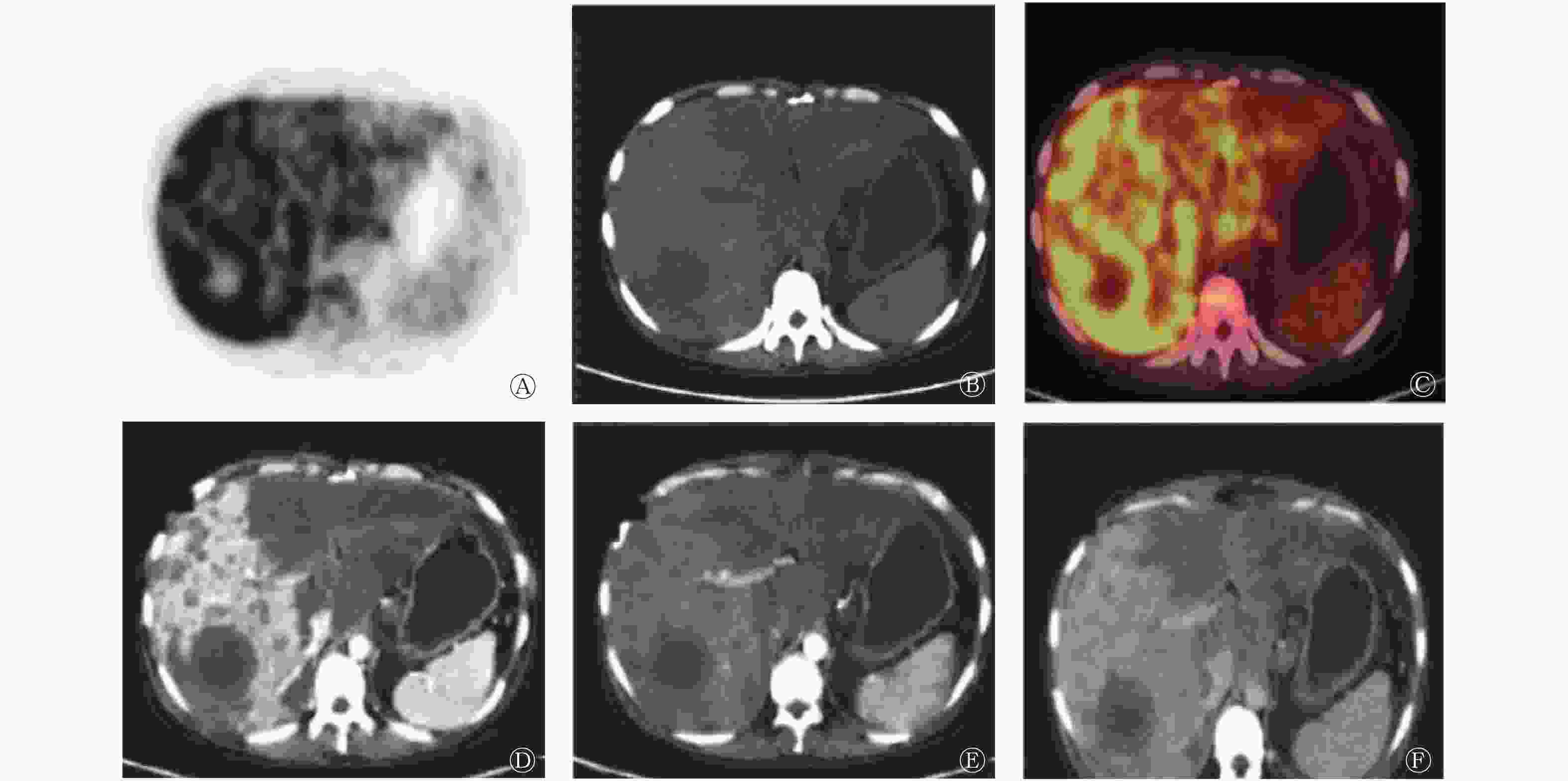
图 1 肝上皮样血管内皮瘤患者(女性,39岁)的18F-FDG PET/CT显像和肝脏Ⅲ期CT增强扫描图 图中,A:PET示肝脏代谢弥漫不均匀增高,局部代谢缺损;B:CT示肝脏体积增大伴密度弥漫不均匀减低,肝右叶示类圆形低密度病灶;C:PET/CT融合图像示肝右叶类圆形病灶中心代谢缺损,且肝左叶密度较低处代谢相对减低。D~F:分别为动脉期、门脉期和延迟期CT增强显像图,示肝右叶大部分呈明显强化,另肝右叶类圆形病灶边缘呈环形低密度,内侧呈环状相对高密度,中心呈低密度,由外到内“低-高-低”呈“靶征”强化,并可见肝右叶粗大血管穿行。FDG: 氟脱氧葡萄糖;PET:正电子发射断层显像术;CT:计算机体层摄影术
Figure 1. 18F-FDG PET/CT imaging and liver triple-phase contrast-enhanced CT images of hepatic epithelioid hemangioendothelioma(female,39 years old)
PET/CT检查4 d后在超声引导下行肝穿刺活检,病理结果:低度恶性肿瘤,结合形态学及免疫组化检测结果,符合HEHE。免疫组化检测结果:CD34(+)、CD31(+)、Vim(+)、S-100(−)、CK5/6(−)、SMA(+)、Actin(+)、Desmin(−)、CD68个别(+)、AACT(+)、CD57(−)、Bcl-2部分(+)、HMB45(−)、MelanA(−)、Hep(−)、Ki-67约15%(+);残存胆管上皮CK(+)、EMA(+)、CK7(+)。
-
HEHE是一种罕见的血管性肝肿瘤,发病原因不明,一般认为与避孕药、肝外伤、肝炎病史、长期接触石棉、氯乙烯或二氧化钍等有关[4]。其临床表现缺乏特异性,部分表现为腹部不适,肝区疼痛,可伴随布加综合征或卡萨巴赫-梅里特综合征,当肿瘤较大时可出现肿瘤破裂并腹腔出血。由于肿瘤早期无症状,该病发现并确诊时大多已是晚期。一般认为根治性手术切除或肝移植是治疗HEHE较好的方法[5-6],该病对放化疗均不敏感,所以往往预后不佳,5年总生存率仅为43%[7]。
HEHE的生物学行为被认为介于血管瘤和血管肉瘤之间,但其病情发展过程及预后往往难以预测,可以进展缓慢,也可以发展很迅速[8]。典型的HEHE在影像学上可有一定特点,CT平扫一般呈多发低密度结节,中心密度更低,部分可见钙化,肿瘤较大时边界模糊;增强扫描可见轻度强化,典型肿瘤可见由外到内呈“低-高-低”环形强化,呈“靶征”改变[9],偶可见粗大的静脉穿行,呈“棒棒糖征”改变[10]。另有文献报道[8],HEHE行18F-FDG PET/CT显像时,代谢呈轻度增高表现。本例患者CT增强扫描可见肝右叶病灶具有典型由外至内“低-高-低”强化的“靶征”表现,中心低密度未见明显强化,PET显像示肝脏葡萄糖代谢弥漫不均匀增高,其中肝右叶病灶中心可见放射性缺损。这与Wang等[11]报道一致,这种代谢特点与其肿瘤学行为及预后的关系尚有待进一步研究。
免疫组化因子CD31、CD34和因子Ⅴ Ⅲ相关抗原阳性更能帮助诊断HEHE。CD31被认为是特异性表达,而CD34具有较高的灵敏度,但不具有特异度[12]。本例患者免疫组化CD31和CD34均呈阳性。
综上所述,HEHE在临床上较为罕见,确诊还需要依靠病理结果,临床上遇到肝脏占位的情况,无法用常见肝脏疾病来解释的时候,应该考虑HEHE,并行组织学检查协助诊断。
利益冲突 本研究由署名作者按以下贡献声明独立开展,不涉及任何利益冲突。
作者贡献声明 孙绮桦负责研究命题的提出、论文的撰写与修订;梁婉媚负责病例图像的分析、文献的搜集;王欣璐负责诊断报告的终审;尹吉林负责研究命题的理论指导;张金赫负责研究命题的提出、论文的审核。
肝上皮样血管内皮细胞瘤18F-FDG PET/CT显像一例
18F-FDG PET/CT imaging of hepatic epithelioid hemangioendothelioma: a case report
-
摘要: 笔者介绍了1例肝上皮样血管内皮细胞瘤18F-FDG PET/CT显像的病例。从临床症状、实验室检查、病理学检查及影像学等方面分析该病的特点。并通过文献复习了解肝上皮样血管内皮细胞瘤的特点。该病发病率极低,早期症状不明显,不易被发现,且临床表现缺乏特异性,难以与常见肝脏肿瘤相鉴别,在临床上易造成误诊。且该病预后欠佳,目前认为最有效的治疗方法是肝移植或根治性手术切除。Abstract: A case of hepatic epithelioid hemangioendothelioma with 18F-FDG PET/CT whole-body imaging was reported. The characteristics of the disease were analyzed from clinical symptoms, laboratory and pathological examination, and imaging, and were understood through literature review. The disease has an extremely low incidence and the clinical manifestations are lack of specificity. It is not easy to be found in the early stage. It is difficult to be distinguished from common liver tumors, and easy to be misdiagnosed. The prognosis of the disease is poor. It is recognized at present that the most effective treatment is liver transplantation or radical resection.
-
Key words:
-
图 1 肝上皮样血管内皮瘤患者(女性,39岁)的18F-FDG PET/CT显像和肝脏Ⅲ期CT增强扫描图 图中,A:PET示肝脏代谢弥漫不均匀增高,局部代谢缺损;B:CT示肝脏体积增大伴密度弥漫不均匀减低,肝右叶示类圆形低密度病灶;C:PET/CT融合图像示肝右叶类圆形病灶中心代谢缺损,且肝左叶密度较低处代谢相对减低。D~F:分别为动脉期、门脉期和延迟期CT增强显像图,示肝右叶大部分呈明显强化,另肝右叶类圆形病灶边缘呈环形低密度,内侧呈环状相对高密度,中心呈低密度,由外到内“低-高-低”呈“靶征”强化,并可见肝右叶粗大血管穿行。FDG: 氟脱氧葡萄糖;PET:正电子发射断层显像术;CT:计算机体层摄影术
Figure 1. 18F-FDG PET/CT imaging and liver triple-phase contrast-enhanced CT images of hepatic epithelioid hemangioendothelioma(female,39 years old)
-
[1] Treska V, Daum O, Svajdler M, et al. Hepatic epithelioid hemangioendothelioma: A rare tumor and diagnostic dilemma[J]. In vivo (Athens, Greece), 2017, 31(4): 763−767. DOI: 10.21873/invivo.11128. [2] Llueca A, Piquer D, Maazouzi Y, et al. Hepatic epithelioid hemangioendothelioma−A great mimicker[J/OL]. Int J Surg Case Rep, 2018, 53: 25−28[2019-12-29]. https://www.ncbi.nlm.nih.gov/pmc/articles/PMC6205146. DOI: 10.1016/j.ijscr.2018.10.013. [3] Manucha V, Sun CC. Cytologic findings and differential diagnosis in hepatic Epithelioid hemangioendothelioma: a case report[J]. Acta cytological, 2008, 52(6): 713−717. DOI: 10.1159/000325627. [4] 孙淑杰, 迮兴宇, 赵新颜. 肝上皮样血管内皮瘤文献复习及临床特点分析[J]. 临床和实验医学杂志, 2012, 11(9): 654−656. DOI: 10.3969/j.issn.1671-4695.2012.09.006.
Sun SJ, Ze XY, Zhao XY. A review of literatures on clinical manifestations of hepatic epithelioid hemagioendothelioma[J]. J Clin Experimental Med, 2012, 11(9): 654−656. DOI: 10.3969/j.issn.1671-4695.2012.09.006.[5] 缪建良, 刘淼, 陈达伟. 肝脏上皮样血管内皮瘤的影像学特征[J]. 放射学实践, 2011, 26(7): 736−738. DOI: 10.3969/j.issn.1000-0313.2011.07.011.
Miu JL, Liu M, Chen DW. Imaging findings of hepatic epithelioid hemangioendothelioma[J]. Radiol Pract, 2011, 26(7): 736−738. DOI: 10.3969/j.issn.1000-0313.2011.07.011.[6] Zhang W, Jambulingam PS, Silva MA, et al. Orthotopic liver transplantation for epithelioid haemangioendothelioma[J]. Eur J Surg Onco, 2007, 33(7): 898−901. DOI: 10.1016/j.ejso.2007.01.021. [7] Makhlouf HR, Ishak KG, Goodman ZD. Epithelioid hemangioendothelioma of the liver: a clinicopathologic study of 137 cases[J]. Cancer, 1999, 85(3): 562−582. DOI: 10.1002/(sici)1097-0142(19990201)85:3<562::aid-cncr7>3.0.co;2-t. [8] Hu HJ, Jin YW, Jing QY, et al. Hepatic epithelioid hemangioendothelioma: Dilemma and challenges in the preoperative diagnosis[J]. World J Gastroenterol, 2016, 22(41): 9247−9250. DOI: 10.3748/wjg.v22.i41.9247. [9] Mamone G, Miraglia R. The "Target sign" and the “Lollipop sign” in hepatic epithelioid hemangioendothelioma[J]. Abdom Radiol (NY), 2019, 44(4): 1617−1620. DOI: 10.1007/s00261-018-1820-9. [10] 徐亚丹, 王文平. 肝上皮样血管内皮瘤影像学表现研究进展[J]. 中华超声影像学杂志, 2016, 25(11): 1008−1011. DOI: 10.3760/cma.j.issn.1004-4477.2016.11.023.
Xu YD, Wang WP. Adcances in imaging diagnosis of hepatic epithelioid hemangioendothelioma[J]. Chin J Ultrasonography, 2016, 25(11): 1008−1011. DOI: 10.3760/cma.j.issn.1004-4477.2016.11.023.[11] Wang WM, Liu GB, Hu PC, et al. Imaging characteristics and prognostic values of hepatic epithelioid hemangioendothelioma on 18F-FDG PET/CT[J]. Clin Exp Med, 2020, 20(4): 557−567. DOI: 10.1007/s10238-020-00653-0. [12] Liu YI, Brown SS, Elihu A, et al. Hepatic epithelioid hemangioendothelioma[J]. Digestive diseases and sciences, 2011, 56(2): 303−306. DOI: 10.1007/s10620-010-1470-4. -

 点击查看大图

图(1)
计量
- 文章访问数: 3109
- HTML全文浏览量: 1968
- PDF下载量: 18