


-
前纵隔是胸骨与气管、主动脉及心包前缘的区域,其内可发生多种恶性肿瘤,且好发于成年人,病理类型多样,影像学表现各异,误诊率较高;同时,由于该区域不同类型恶性肿瘤的治疗方法各异,因此早期诊断对该病的临床分期、治疗及预后具有重要意义[1-2]。虽然以往文献中有关于该部位肿瘤的影像学诊断的报道,但有关18F-FDG PET/CT与增强CT在成人原发前纵隔恶性肿瘤中的研究报道较为少见。本研究比较上述2种显像方法在成人原发前纵隔恶性肿瘤中的诊断效能,以提高临床上对该部位病变的诊断水平。
-
回顾性分析2016年6月至2019年5月在湖北文理学院附属医院(襄阳市中心医院)经病理证实的成人原发前纵隔肿瘤患者80例,其中,恶性肿瘤35例、良性肿瘤45例;男性46例、女性34例,年龄20~80(45.5±10.2)岁。纳入标准:(1)患者年龄≥18岁;(2)肿块位于前纵隔内,且检查前未经病理确诊;(3)无其他恶性肿瘤病史。排除标准:(1)患者年龄<18岁;(2)肿块位于纵隔其他位置,或肺、食管或胸骨等肿瘤累及前纵隔;(3)伴有其他恶性肿瘤或恶性肿瘤史。所有患者或其家属均签署了知情同意书,本研究获得了湖北文理学院附属医院(襄阳市中心医院)伦理委员会的批准(批准号:2019-078)。
-
CT扫描使用德国Siemens公司 Sensation 16 CT仪和荷兰 Philips公司Brilliance 64 CT仪,80例患者均行胸部CT平扫+增强扫描,扫描范围从肺尖至膈肌下,增强扫描使用高压注射器经肘静脉注射江苏恒瑞医药股份有限公司对比剂碘佛醇(320 mgI/mL),注射速度为3 mL/s,剂量为1.0~1.5 mL/kg。PET/CT显像使用中国联影公司uMI510 PET/CT仪,检查前患者空腹6 h以上,血糖控制在7.0 mmol/L以内,静脉注射由美国GE公司回旋加速器生产的18F-FDG,放射化学纯度≥95%,剂量为3.70~7.40 MBq/kg,平卧休息1 h后行全身扫描。2种检查的间隔时间在2周内,所有扫描数据均进行多平面重建后处理,再由2名具有10年以上工作经验的影像医师共同阅片得出诊断结果,意见不一致时以高年资医师意见为准。最终结果与术后病理学检查或穿刺活检病理学及免疫组织化学结果进行比较,分别计算并比较2种检查方法对成人原发前纵隔恶性肿瘤诊断的灵敏度、特异度、准确率,以及对不同类型恶性肿瘤的诊断效能。
-
采用SPSS 21.0软件进行统计学分析。计量资料符合正态分布,以
$\bar x $ ±s表示,18F-FDG PET/CT与增强CT对成人原发前纵隔恶性肿瘤诊断的灵敏度、特异度、准确率的比较,以及对不同类型恶性肿瘤的诊断效能的比较均采用连续性校正χ2检验。检验水准a=0.05,P<0.05为差异有统计学意义。 -
35例恶性肿瘤中,胸腺癌15例(鳞癌8例、腺癌4例、未分化癌3例)(图1中A~C),淋巴瘤10例(非霍奇金淋巴瘤7例、霍奇金淋巴瘤3例)(图1中D~F),恶性生殖细胞瘤7例(精原细胞瘤3例、未成熟畸胎瘤3例、卵黄囊瘤1例)(图1中G~I),神经内分泌癌(非典型类癌)、恶性黑色素瘤及滑膜肉瘤各1例(表1)。
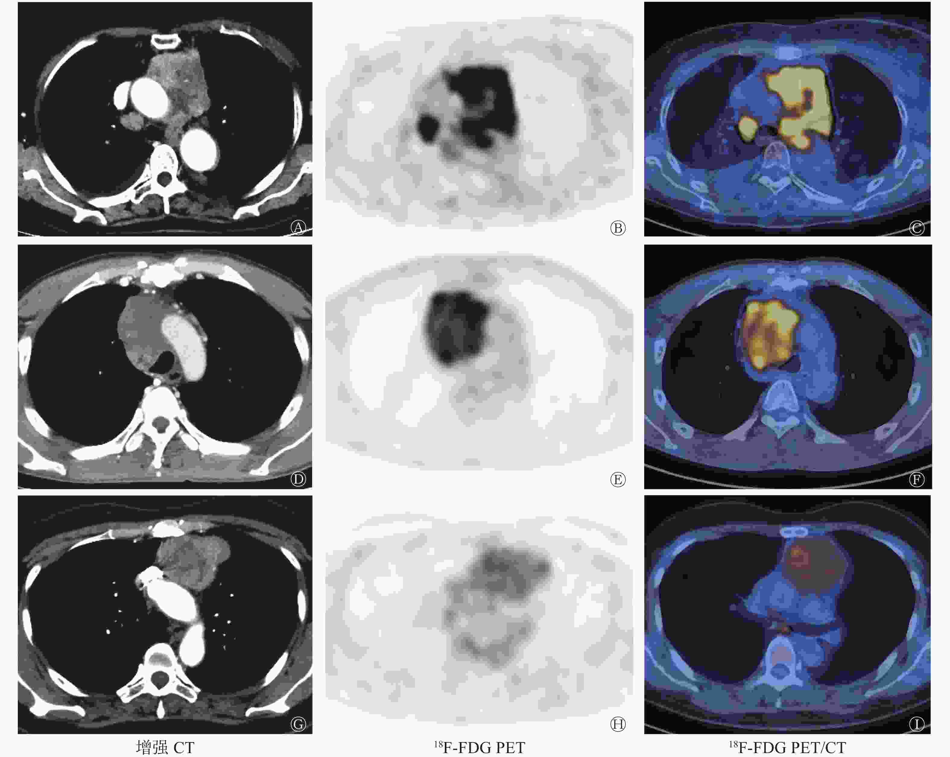
图 1 成人原发前纵隔恶性肿瘤的增强CT与18F-FDG PET/CT显像图
Figure 1. Enhanced CT and 18F-FDG PET/CT for adult patients with primary malignant tumors of the anterior mediastinum
肿瘤类型 例数 男性/女性(例) 年龄(岁) 胸腺癌 15 11/4 55.3±5.2 淋巴瘤 10 4/6 30.5±4.3 恶性生殖细胞瘤 7 4/3 25.7±3.5 神经内分泌癌 1 0/1 65 恶性黑色素瘤 1 0/1 54 滑膜肉瘤 1 0/1 51 表 1 35例成人原发前纵隔恶性肿瘤的一般临床情况
Table 1. General clinical data of 35 adult patients with primary malignant tumors of the anterior mediastinum
-
18F-FDG PET/CT诊断成人原发前纵隔恶性肿瘤的灵敏度、特异度、准确率分别为97.1% (34/35)、93.3% (42/45)、95.0% (76/80),其中,1例神经内分泌癌漏诊为良性病变,2例胸腺瘤及1例淋巴结结核误诊为恶性病变;增强CT诊断的灵敏度、特异度、准确率分别为71.4% (25/35)、77.8% (35/45)、75.0% (60/80),其中,4例胸腺癌漏诊为胸腺瘤,3例恶性生殖细胞瘤、1例淋巴瘤、1例神经内分泌癌及1例滑膜肉瘤均漏诊为良性病变,5例胸腺瘤误诊为胸腺癌,2例结节病误诊为淋巴瘤,2例良性生殖细胞瘤及1例胸腺脓肿误诊为恶性病变。18F-FDG PET/CT对成人原发前纵隔恶性肿瘤诊断的灵敏度、特异度及准确率均高于增强CT,且差异均有统计学意义(χ2=8.612、4.357、12.471,均P<0.05)(表2)。
检查方法 例数 灵敏度 特异度 准确率 真阳性 假阴性 真阴性 假阳性 准确诊断 错误诊断 18F-FDG PET/CT 80 34 1 42 3 76 4 增强CT 80 25 10 35 10 60 20 χ2值 8.612 4.357 12.471 P值 <0.05 <0.05 <0.05 注:表中,FDG:氟脱氧葡萄糖;PET:正电子发射断层显像术;CT:计算机体层摄影术 表 2 18F-FDG PET/CT与增强CT对成人原发前纵隔恶性肿瘤诊断效能的比较(例)
Table 2. Comparison of the sensitivity, specificity and accuracy of 18F-FDG PET/CT and enhanced CT examination for the diagnosis of adult patients with primary malignant tumors of the anterior mediastinum (case)
-
18F-FDG PET/CT对胸腺癌、淋巴瘤及恶性生殖细胞瘤诊断的准确率分别为93.3%(14/15)、100.0%(10/10)、85.7%(6/7);增强CT诊断的准确率分别为60.0%(9/15)、50.0%(5/10)、28.6%(2/7),前者诊断的准确率均高于后者,且差异均有统计学意义(χ2=4.503、6.333、4.333,均P<0.05)。
-
原发于前纵隔的恶性肿瘤种类繁多,约占所有纵隔恶性肿瘤的50%,以成年人多见,其组织学来源多样,主要包括胸腺上皮、淋巴组织、生殖细胞、间叶组织及其他组织来源等,其中以胸腺上皮来源为主[3-4]。本研究中胸腺癌、淋巴瘤及恶性生殖细胞瘤患者较多。基于前纵隔的解剖学特征及位于其内恶性肿瘤的组织学特点,该类肿瘤的恶性程度高,生长速度快,肿块常压迫、侵犯邻近纵隔组织结构或远处转移引起相应症状[4-5]。同时,该部位不同类型恶性肿瘤的治疗方法各异,如胸腺癌、神经内分泌癌、恶性黑色素瘤及滑膜肉瘤等早期常以外科手术切除治疗为主,淋巴瘤以全身化疗为主,而恶性生殖细胞瘤则在行外科手术治疗前常需进行全身化疗[1,6-8]。因此,成人原发前纵隔恶性肿瘤的早期诊断具有重要的临床意义,可指导临床分期、治疗方案的选择及进行预后评估,最终提高患者的生存率及生活质量。
目前,成人原发前纵隔恶性肿瘤的影像学检查方法主要包括胸部CT平扫、增强CT及18F-FDG PET/CT。胸部CT平扫仅能大致显示病灶的位置、范围及内部高密度出血、钙化等情况,不易与周围结构进行区分,难以作出定性诊断,常用于临床初步筛查。而增强CT检查能够更加清晰准确地显示病灶的形态、密度、血供及与周围组织的关系,有助于对部分肿瘤作出较为正确的定性诊断,可作为CT平扫后的进一步检查,但对于局部侵犯及转移灶的诊断及评估的价值相对有限[9]。18F-FDG PET/CT显像是从分子生物学的角度进行功能成像,能反映组织细胞内的代谢情况,且能提供较为精确的解剖定位,其一次扫描不仅可了解全身情况,还可通过测量半定量指标(SUVmax)来反映肿瘤细胞对18F-FDG的摄取情况,从而显示肿瘤的生长代谢活性,加之其准确的定位,能更早地发现一些较小的转移灶,有助于疾病的诊断及鉴别诊断,更加有利于指导临床分期、治疗及疗效评估[10-11],还能为病灶病检组织学取材提供依据,避免不必要的穿刺活检。
原发于成人的前纵隔恶性肿瘤种类繁多,侵袭性高,影像学表现多样,导致其误诊率较高,且不同类型恶性肿瘤治疗方法不同,因此早期诊断十分重要。本研究结果显示,18F-FDG PET/CT诊断成人原发前纵隔恶性肿瘤的灵敏度、特异度及准确率均高于增强CT,在35例恶性肿瘤中仅有1例恶性神经内分泌癌因病灶呈环形浓聚,糖代谢轻度增高,漏诊为良性病变;在45例良性肿瘤中有3例(2例胸腺瘤及1例淋巴结结核)因病灶边界不清伴纵隔淋巴结糖代谢异常增高,误诊为恶性病变;总体来看18F-FDG PET/CT的诊断准确率较高[95%(76/80)]。增强CT漏诊及误诊各10例,其中10例恶性肿瘤因忽视了病灶的局部侵犯和(或)远处转移等情况,漏诊为良性病变;5例胸腺瘤因病灶体积较大且内部囊变坏死明显,误诊为胸腺癌;2例结节病因前纵隔病变伴纵隔多发淋巴结肿大,误诊为淋巴瘤;2例纵隔良性生殖细胞瘤因病灶体积较大、形态不规则且内部缺乏脂肪成分,误诊为恶性生殖源性肿瘤;1例胸腺脓肿因病灶边界不清,其内坏死明显且伴纵隔淋巴结肿大,误诊为胸腺癌;总体来看增强CT的诊断准确率偏低[75%(60/80)]。增强CT的诊断准确率低于18F-FDG PET/CT的原因可能是由于18F-FDG PET/CT反映的是恶性肿瘤内癌细胞的葡萄糖代谢情况,其摄取增多与癌细胞迅速生长繁殖所需要的能量增加有关,而增强CT主要反映的是肿瘤内部的供血情况,这与文献[1,12]报道相符。对于前纵隔内不同类型恶性肿瘤的诊断准确率,本研究结果显示18F-FDG PET/CT诊断胸腺癌、淋巴瘤及恶性生殖细胞瘤的准确率均高于增强CT,因为18F-FDG PET/CT进行的是功能成像,可反映病灶分子生物学水平的变化,其对于局部侵犯及转移灶的诊断及评估优于增强CT,再结合患者的临床资料更加有助于病灶的诊断,尤其是对于前纵隔淋巴瘤的诊断,18F-FDG PET/CT能发现除前纵隔外增强CT无法发现的更多的病灶,且更加有助于病检取材部位的选择。值得注意的是,本研究中增强CT对于恶性生殖细胞瘤诊断的准确率明显低于18F-FDG PET/CT,可能与影像科医师忽视了人绒毛膜促性腺激素(β-HCG)和甲胎蛋白(AFP)等实验室检查有关。本研究中有6例恶性生殖细胞瘤伴人绒毛膜促性腺激素(β-HCG)和(或)甲胎蛋白(AFP)水平不同程度的升高,这与文献[13-14]报道的纵隔恶性生殖细胞瘤常伴人绒毛膜促性腺激素(β-HCG)、甲胎蛋白(AFP)水平升高相符。而对神经内分泌癌、恶性黑色素瘤及滑膜肉瘤的诊断,因这3种疾病病例数较少不能够进行统计学分析,今后需增大样本量进行进一步研究。我们还发现胸腺癌肿瘤体积大多小于淋巴瘤和恶性生殖细胞瘤,这可能与胸腺癌细胞基质缺乏且多形性明显,癌细胞生长迅速更易局部侵犯引起相应临床症状导致患者就诊较早有关。本研究中淋巴瘤的18F-FDG摄取最高,SUVmax均值约达20.1,而胸腺癌和恶性生殖细胞瘤的SUVmax均值约为15.4和13.6,这与文献[1,10,12]报道相符。此外,本研究中淋巴瘤和滑膜肉瘤包绕邻近纵隔血管生长,增强CT扫描后胸腺癌、恶性生殖细胞瘤和滑膜肉瘤呈明显不均匀强化,淋巴瘤呈轻中度强化,神经内分泌癌呈明显环形强化并可见强化分隔,恶性黑色素瘤呈轻度不均匀强化。以上影像学特点可能有助于成人原发前纵隔恶性肿瘤的诊断及鉴别诊断,但对于指导临床分期18F-FDG PET/CT仍优于增强CT。
综上所述,18F-FDG PET/CT能够从分子水平反映成人原发前纵隔恶性肿瘤的代谢情况,具有重要的诊断价值,其诊断效能优于增强CT,可作为成人原发前纵隔恶性肿瘤的主要检查方法。
利益冲突 本研究由署名作者按以下贡献声明独立开展,不涉及任何利益冲突。
作者贡献声明 陈涛负责命题的提出与设计、论文的撰写;樊建中负责图像的采集与分析;李文菲、吴彩云负责论文的修订与审校、数据的统计与分析。
18F-FDG PET/CT与增强CT在成人原发前纵隔恶性肿瘤中的诊断价值
Diagnostic value of 18F-FDG PET/CT and enhanced CT for adult patients with primary malignant tumors of the anterior mediastinum
-
摘要:
目的 探讨18F-氟脱氧葡萄糖(FDG) PET/CT与增强CT在成人原发前纵隔恶性肿瘤中的诊断价值,以提高对该部位病变的诊断水平。 方法 回顾性分析2016年6月至2019年5月在湖北文理学院附属医院(襄阳市中心医院)经病理证实的成人原发前纵隔肿瘤患者80例(恶性肿瘤35例、良性肿瘤45例),其中,男性46例、女性34例,年龄20~80(45.5±10.2)岁。所有患者均行全身18F-FDG PET/CT及胸部CT平扫+增强扫描,2种检查的间隔时间在2周内。分别计算并采用χ2检验比较18F-FDG PET/CT与增强CT扫描的诊断灵敏度、特异度和准确率,采用χ2检验比较2种检查方法对不同类型恶性肿瘤的诊断效能。 结果 35例恶性肿瘤中胸腺癌15例,淋巴瘤10例,恶性生殖细胞瘤7例,神经内分泌癌、恶性黑色素瘤及滑膜肉瘤各1例。18F-FDG PET/CT诊断成人原发前纵隔恶性肿瘤的灵敏度[97.1%(34/35)]、特异度[93.3%(42/45)]及准确率[95.0%(76/80)]均高于增强CT[71.4%(25/35)、77.8%(35/45)、75.0%(60/80)],且差异有统计学意义(χ2=8.612、4.357、12.471,均P<0.05);18F-FDG PET/CT对胸腺癌、淋巴瘤及恶性生殖细胞瘤诊断的准确率[93.3%(14/15)、100.0%(10/10)、85.7%(6/7)]均高于增强CT[60.0%(9/15)、50.0%(5/10)、28.6%(2/7)],且差异有统计学意义(χ2=4.503、6.333、4.333,均P<0.05)。 结论 18F-FDG PET/CT对成人原发前纵隔恶性肿瘤具有重要的诊断价值,其诊断效能优于增强CT,可作为该病主要的检查方法。 -
关键词:
- 肿瘤 /
- 正电子发射断层显像术 /
- 体层摄影术, X线计算机 /
- 氟脱氧葡萄糖F18 /
- 前纵隔
Abstract:Objective To compare the diagnostic value of 18F-fluorodeoxyglucose (FDG) PET/CT versus enhanced CT in detecting adult patients with primary malignant tumors of the anterior mediastinum and improve the accuracy of diagnosis. Methods A total of 80 adult patients with primary tumors of the anterior mediastinum (35 malignant tumors and 45 benign tumors) confirmed by pathology from June 2016 to May 2019 in Affiliated Hospital of Hubei University of Arts and Science (Xiangyang Central Hospital) were retrospectively analyzed. The patients included 46 males and 34 females aged 20–80 years, with a median age of 45.5±10.2 years. All patients were subjected to whole-body 18F-FDG PET/CT and plain and enhanced-scan thorax CT within a 2-week interval. The sensitivity, specificity, and accuracy of 18F-FDG PET/CT and enhanced CT for the diagnosis of adult patients with primary malignant tumors of the anterior mediastinum were calculated and compared by χ2 test. The diagnostic efficiency of 18F-FDG PET/CT versus enhanced CT for different types of adult patients with primary malignant tumors of the anterior mediastinum was also compared by χ2 test. Results Among the 35 malignant tumors, 15 were thymic carcinomas, 10 were lymphomas, 7 were malignant germinomas, 1 was neuroendocrine carcinoma, 1 was malignant melanoma, and 1 was synovial sarcoma. The sensitivity, specificity, and accuracy [97.1%(34/35), 93.3%(42/45), 95.0%(76/80), respectively] of 18F-FDG PET/CT for the diagnosis of adult patients with primary malignant tumors of the anterior mediastinum were significantly higher than those of enhanced CT [71.4%(25/35), 77.8%(35/45), 75.0%(60/80), respectively; χ2=8.612, 4.357, 12.471, respectively; all P<0.05]. The diagnostic accuracies of 18F-FDG PET/CT for thymic carcinoma, lymphoma, and malignant germinoma [93.3%(14/15), 100.0%(10/10), 85.7%(6/7), respectively] were significantly higher than those of enhanced CT [60.0%(9/15), 50.0%(5/10), 28.6%(2/7), respectively; χ2=4.503, 6.333, 4.333, respectively; all P<0.05]. Conclusions 18F-FDG PET/CT has significant diagnostic value for adult patients with primary malignant tumors of the anterior mediastinum. The diagnostic efficiency of this technique is better than that of enhanced CT, and it can be used as a main imaging method for the diagnosis of these types of diseases. -
图 1 成人原发前纵隔恶性肿瘤的增强CT与18F-FDG PET/CT显像图
Figure 1. Enhanced CT and 18F-FDG PET/CT for adult patients with primary malignant tumors of the anterior mediastinum
表 1 35例成人原发前纵隔恶性肿瘤的一般临床情况
Table 1. General clinical data of 35 adult patients with primary malignant tumors of the anterior mediastinum
肿瘤类型 例数 男性/女性(例) 年龄(岁) 胸腺癌 15 11/4 55.3±5.2 淋巴瘤 10 4/6 30.5±4.3 恶性生殖细胞瘤 7 4/3 25.7±3.5 神经内分泌癌 1 0/1 65 恶性黑色素瘤 1 0/1 54 滑膜肉瘤 1 0/1 51  下载: 导出CSV
表 2 18F-FDG PET/CT与增强CT对成人原发前纵隔恶性肿瘤诊断效能的比较(例)
Table 2. Comparison of the sensitivity, specificity and accuracy of 18F-FDG PET/CT and enhanced CT examination for the diagnosis of adult patients with primary malignant tumors of the anterior mediastinum (case)
检查方法 例数 灵敏度 特异度 准确率 真阳性 假阴性 真阴性 假阳性 准确诊断 错误诊断 18F-FDG PET/CT 80 34 1 42 3 76 4 增强CT 80 25 10 35 10 60 20 χ2值 8.612 4.357 12.471 P值 <0.05 <0.05 <0.05 注:表中,FDG:氟脱氧葡萄糖;PET:正电子发射断层显像术;CT:计算机体层摄影术
下载: 导出CSV
-
[1] Yabuuchi H, Matsuo Y, Abe K, et al. Anterior mediastinal solid tumours in adults: characterisation using dynamic contrast-enhanced MRI, diffusion-weighted MRI, and FDG-PET/CT[J]. Clin Radiol, 2015, 70(11): 1289−1298. DOI: 10.1016/j.crad.2015.07.004. [2] Ocal M, Yildiz B, Karadurmus N, et al. Comparison of the clinical features and hematopoietic stem cell transplantation outcomes of mediastinal malignant germ cell tumors with nonmediastinal extragonadal placements[J/OL]. Onco Targets Ther, 2016, 9(5): 7445−7450[2019-06-06]. https://www.ncbi.nlm.nih.gov/pmc/journals/1214. DOI: 10.2147/OTT.S107899. [3] Malik R, Mullassery D, Kleine-Brueggeney M, et al. Anterior mediastinal masses—a multidisciplinary pathway for safe diagnostic procedures[J]. J Pediatr Surg, 2019, 54(2): 251−254. DOI: 10.1016/j.jpedsurg.2018.10.080. [4] Hakiri S, Kawaguchi K, Fukui T, et al. Verification of the diagnostic strategy for anterior mediastinal tumors[J]. Int J Clin Oncol, 2019, 24(4): 385−393. DOI: 10.1007/s10147-018-1362-8. [5] Watanabe T, Shimomura H, Mutoh T, et al. Positron emission tomography/computed tomography as a clinical diagnostic tool for anterior mediastinal tumors[J]. Surg Today, 2019, 49(2): 143−149. DOI: 10.1007/s00595-018-1712-1. [6] Youn HC. A primary malignant melanoma of the mediastinum with gross surgical view[J]. J Thorac Dis, 2016, 8(1): E133−E136. DOI: 10.3978/j.issn.2072-1439.2016.01.22. [7] Abu-Zaid A, AlNajjar A, Alotaibi S, et al. Huge primary mediastinal synovial sarcoma fully occupying the right hemithorax[J]. J Cancer Res Ther, 2018, 14(3): 682−686. DOI: 10.4103/0973-1482.172137. [8] Nakhla SG, Sundararajan S. A rare case of primary anterior mediastinal yolk sac tumor in an elderly adult male[J/OL]. Case Rep Oncol Med, 2016, 2016: 8961486[2019-06-06]. https://www.ncbi.nlm.nih.gov/pmc/articles/PMC4837257. DOI: 10.1155/2016/8961486. [9] Hammer MM, Miskin N, Madan R, et al. Predictive features for anterior mediastinal mass diagnoses[J]. J Comput Assist Tomogr, 2019, 43(1): 98−103. DOI: 10.1097/RCT.0000000000000782. [10] Ceriani L, Milan L, Martelli M, et al. Metabolic heterogeneity on baseline 18FDG-PET/CT scan is a predictor of outcome in primary mediastinal B-cell lymphoma[J]. Blood, 2018, 132(2): 179−186. DOI: 10.1182/blood-2018-01-826958. [11] Tatci E, Ozmen O, Dadali Y, et al. The role of FDG PET/CT in evaluation of mediastinal masses and neurogenic tumors of chest wall[J/OL]. Int J Clin Exp Med, 2015, 8(7): 11146−11152[2019-06-06]. http://www.ncbi.nlm.nih.gov/pmc/articles/PMC4565299. [12] 丁重阳, 李天女. 前纵隔肿瘤18F-FDG PET/CT显像特征[J]. 中国医学影像技术, 2014, 30(4): 531−534. DOI: 10.13929/j.1003-3289.2014.04.010.
Ding CY, Li TN. Features of anterior mediastinal tumors in 18F-FDG PET/CT imaging[J]. Chin J Med Imaging Technol, 2014, 30(4): 531−534. DOI: 10.13929/j.1003-3289.2014.04.010.[13] 陈涛, 严静东, 雷贞妮. 前纵隔原发性恶性肿瘤的MSCT影像表现[J]. 实用医学杂志, 2015, 31(18): 3039−3042. DOI: 10.3969/j.issn.1006-5725.2015.18.033.
Chen Tao, Yan JD, Lei ZN. MSCT imaging findings in anterior mediastinal primary malignant tumors[J]. J Pract Med, 2015, 31(18): 3039−3042. DOI: 10.3969/j.issn.1006-5725.2015.18.033.[14] Joly C, Deblock M, Desandes E, et al. Primary mediastinal germs cells tumors: a twenty years experience in a comprehensive cancer center[J]. Bull Cancer, 2014, 101(12): 1067−1073. DOI: 10.1684/bdc.2014.2047. -

 点击查看大图

图(1)表(2)
计量
- 文章访问数: 3058
- HTML全文浏览量: 2221
- PDF下载量: 15
