


 下载:
下载:
-
异位胰腺亦称迷走胰腺,是指生长在其他部位的孤立的胰腺组织,与正常胰腺组织无解剖上的联系,属于先天性畸形[1]。异位胰腺作为一种罕见的组织异位病变,在临床上多为偶然发现,尸检发现率为0.6%~13.7%[2]。异位胰腺患者通常无明显临床症状及体征,一旦出现并发症,则常表现为腹痛、消化道出血、黄疸等[3]。由于缺乏特异性标志物和检测方法的局限性,临床上难以通过实验室检查和常规影像学检查来准确诊断[4-5]。异位胰腺组织多分布于消化道,其中肝脏异位胰腺更为罕见。笔者通过分析该病例的18F-FDG PET/CT影像学表现及对相关文献进行复习,以加深对肝脏异位胰腺影像学特征的认识。
-
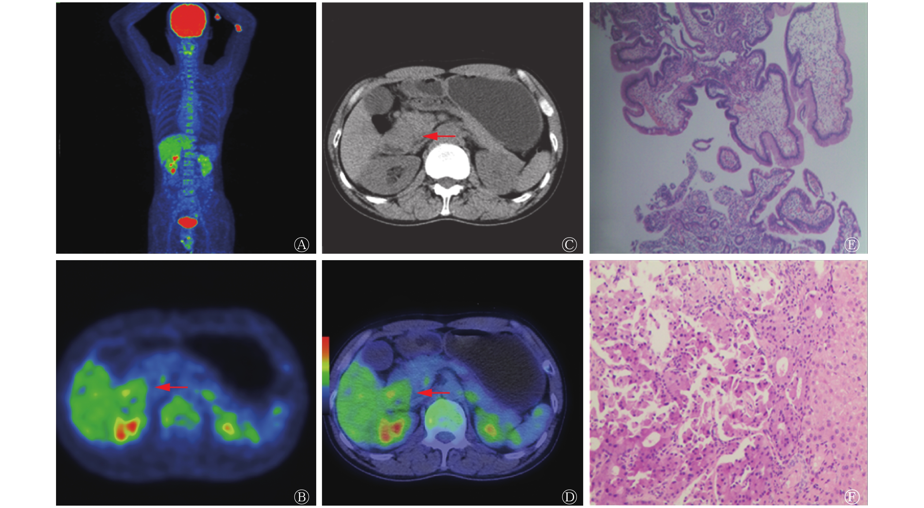
患者男性,45岁,因“腹痛、腹胀1年,病情加重半月”入院。体格检查未见明显腹部触痛、按压痛及叩击痛,未扪及明显腹部肿块。实验室检查结果:WBC 5.59×10⁹个/L(正常值:3.5×109~9.5×109个/L)、血红蛋白163.00 g/L(正常值:130~175 g/L)、血小板286.00×109个/L(正常值:125×109~350×109个/L)、嗜中性粒细胞73.2%(正常值:40%~75%)。生化检查结果:丙氨酸氨基转移酶13 U/L(正常值:9~50 U/L)、门冬氨酸氨基转移酶18 U/L(正常值:15~40 U/L)、肌酐78 μmol/L(正常值:32~106 μmol/L)。肿瘤3项结果:糖类抗原199为7.63 U/mL(正常值:0~37 U/mL)、甲胎蛋白2.4 ng/mL(正常值:0~12 ng/mL)、癌胚抗原2.48 ng/mL(正常值:0~7 ng/mL)。凝血4项未见明显异常阳性指标。为明确诊断,进一步对患者行影像学检查。超声内镜检查示十二指肠降段不均匀低回声肿块。胃镜病理结果示十二指肠降段肿块组织固有层存在大量淋巴细胞、嗜酸性粒细胞、浆细胞浸润。18F-FDG PET(美国GE公司的Discovery Elite 690)显像(图1中A~D)示:十二指肠降段局部见一类圆形肿块,18F-FDG摄取异常增高,SUVmax为3.0,肝脏未见明显18F-FDG异常分布;同机CT显示:十二指肠降段管腔变窄,管壁增厚,局部见一类圆形肿块,大小约2.8 cm×2.6 cm×3.0 cm,密度均匀,CT值约47 HU;肝脏形态正常,未见明显异常密度影。PET/CT检查结果提示:十二指肠降段肿块,葡萄糖代谢增高,考虑小肠间质瘤可能性大。患者随后行手术治疗,术中可见十二指肠降段有一大小约为3 cm×4 cm肿块,质硬,活动度差,未突破浆膜层;在肝脏中可见大量的粟粒状结节,直径为1~3 mm。术后分别从十二指肠肿块和肝脏中取活组织进行病理诊断,结果:肝内异位胰腺(图1中E),十二指肠组织炎性改变(图1中F)。患者术后接受拉氧头孢钠、生长抑素抗感染治疗,症状改善后出院。
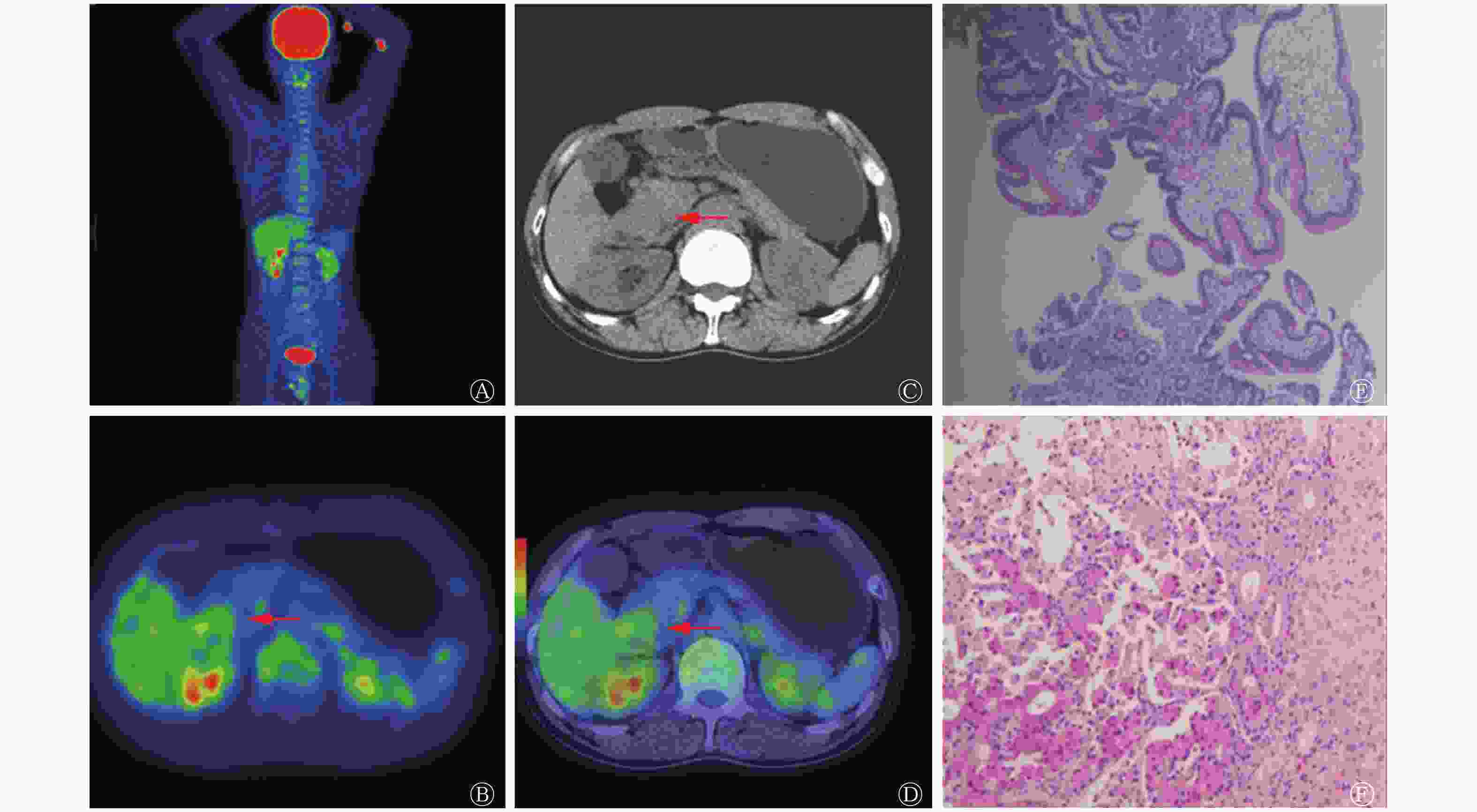
图 1 肝内异位胰腺患者(男性,45岁)的18F-FDG PET/CT显像图及病理图(苏木精-伊红染色,×40)
Figure 1. 18F-FDG PET/CT imaging and pathological map in patients(male,45 years old)with hepatic ectopic pancreas
-
异位胰腺为异常部位的胰腺组织,与正常胰腺无解剖、神经或血管连接,是胚胎发育过程中形成的一种先天性畸形。其可能的原因是胰腺原基与胚胎原肠黏连或穿透原肠壁,并随原肠旋转而分布于各个异常的部位,也是内胚层异向分化的结果[6-7]。异位胰腺组织可位于消化道的任何位置,常见部位为胃、十二指肠、空肠、回肠及Meckel憩室,结肠、胆囊、胆管、肝脏、脾脏、盆腔等部位则相对少见[2-3, 8-9]。
异位胰腺属于局部异常组织,会受到正常组织的排异反应;且异位胰腺具有外分泌功能,其分泌的胰蛋白酶等分泌物会对消化道器官造成刺激,进而引起一系列病理变化。因此,异位胰腺可引起多种并发症,包括炎症、溃疡、出血、外分泌囊肿、胰瘘以及恶变(腺癌),还可引起幽门梗阻、胆管梗阻、肠套叠、肠梗阻等症状[5, 10]。然而,异位胰腺患者临床表现无特异性,这与其发生部位、大小和并发症有关[11]。本例肝脏异位胰腺患者的主要临床症状为腹痛、腹胀,其体格检查和实验室检查结果未能提供有价值的诊断信息,影像学检查的主要阳性病灶为肝脏异位胰腺并发十二指肠降段炎症病变,需要行病理学检查以明确诊断。
已报道的肝脏异位胰腺常见于肝胆管内,肝实质内少见[12-15]。肝胆管内异位胰腺病变在超声、内镜下逆行性胰胆管造影术显像中的主要表现为肝胆管狭窄、阻塞、扩张和囊变。在CT及MRI中还可以观察到病灶本身的形状、大小、CT密度、MRI信号及强化方式等影像学特征[16-20],CT上可表现为囊性或实性结节,囊性病灶呈囊壁强化,实性病灶强化方式以动脉期明显强化、门脉期缓慢下降为主,这一点与正常部位的胰腺组织强化方式相仿;磁共振胰胆管造影可见胆管扩张或充盈缺损,MRI平扫信号与胰腺实质相近,增强扫描明显不均匀强化。肝实质内异位胰腺病变主要表现为孤立性肿块、囊肿和弥漫性小结节[21-22]。
随着18F-FDG PET/CT检查时偶然发现消化道异位胰腺的病例增多,其诊断异位胰腺的应用价值也逐渐受到关注。目前已有多项消化道异位胰腺18F-FDG PET/CT显像的报道[2-4, 6-7],但未检索到肝脏异位胰腺的18F-FDG PET/CT显像报道。由于异位胰腺的占位效应和分泌功能,其原发灶及并发灶在18F-FDG PET/CT显像中往往表现为局部肿块并伴有18F-FDG的异常摄取[23-27]。本病例中,肝脏异位胰腺病灶在18F-FDG PET/CT显像中未见明显的形态学改变和葡萄糖代谢异常,其可能的原因有以下几点:①异位胰腺组织体积过小,密度与肝实质相近,仅凭肉眼观察难以鉴别;②肝实质18F-FDG本底摄取高,掩盖了异位胰腺的18F-FDG显像;③PET/CT组织分辨率较低,对微小病灶具有局限性。但本病例肝脏异位胰腺的十二指肠炎症并发灶有明显阳性征象,表现为软组织肿块合并18F-FDG异常摄取增高,这一结果表明18F-FDG PET/CT对异位胰腺相关的组织结构和功能变化具有较灵敏的检出效能。18F-FDG PET/CT显像能够为临床诊断异位胰腺提供影像支持,其肝脏异位胰腺病变的表现与病灶的部位、大小和并发症相关。
肝脏异位胰腺合并十二指肠炎症临床十分罕见,本病例中肝脏弥漫性小结节仅在术中可见而无阳性的影像学征象,18F-FDG PET/CT显像中的主要病灶为十二指肠降段肿块,其具有边缘光滑、密度均匀、18F-FDG异常摄取和可疑多发转移的交界性影像及临床特征,与原发性十二指肠良恶性肿瘤的鉴别诊断存在一定困难[28]。尽管临床上胃肠道间质瘤的恶性比例只有10%~30%,但所有胃肠道间质瘤均有恶性潜能[29],结合本病例特点,故考虑十二指肠间质瘤的可能性大。但最后病理结果证实为肝脏异位胰腺合并十二指肠炎症,提示今后在鉴别诊断十二指肠肿瘤病变时,当病变具有上述交界性影像及临床特征时,还应考虑异位胰腺的可能。本病例在18F-FDG PET/CT显像中肝内异位胰腺病灶表现阴性征象,而十二指肠并发炎症病灶表现阳性征象,这对认识肝脏异位胰腺病灶的直接影像特征存在不足,但仍可为增进医务工作者对肝脏异位胰腺影像特征的了解提供素材,为鉴别诊断十二指肠占位性病变提供新的思路。
利益冲突 本研究由署名作者按以下贡献声明独立开展,不涉及任何利益冲突。
作者贡献声明 麦锦慈负责研究命题的设计与论文的撰写;谭志强、唐勇进负责资料的收集;凌雪英负责论文的修改;徐浩负责论文的审阅。
肝脏异位胰腺18F-FDG PET/CT影像学表现一例
18F-FDG PET/CT imaging of hepatic ectopic pancreas: a case report
-
摘要: 异位胰腺多发生在消化道,肝脏异位胰腺临床非常罕见。笔者报道了1例行18F-FDG PET/CT检查且经手术病理结果证实的肝脏异位胰腺病例,其影像学特征主要表现为十二指肠肿块并伴有18F-FDG异常摄取,考虑为与肝脏异位胰腺相关的炎性病灶表现。笔者拟通过对该病例进行分析并对相关文献进行复习,以加深对肝脏异位胰腺影像学特征的认识。Abstract: Ectopic pancreas often occurs in the digestive tract, and hepatic ectopic pancreas syndrome is very rare clinically. This article reports a case of hepatic ectopic pancreas detected by 18F-FDG PET/CT and confirmed by surgery and pathology. The imaging features were duodenal mass with abnormal uptake of 18F-FDG, which was considered to be an inflammatory lesion associated with hepatic ectopic pancreas. By analyzing this case and reviewing the relevant literature, the writer intends to deepens the understanding of the imaging features of hepatic ectopic pancreas through literature review.
-
Key words:
-
[1] Flores A, Papafragkakis C, Uberoi AS, et al. EUS of an atypical ectopic pancreas[J]. Endosc Ultrasound, 2018, 7(3): 216−217. DOI: 10.4103/eus.eus_111_17. [2] Chang TK, Huang CW, Ma CJ, et al. Ectopic pancreas mimicking gastric submucosal tumour treated using robotic surgery[J]. J Minim Access Surg, 2019, 16(2): 179−181. DOI: 10.4103/JMAS_1_19. [3] Xiang SH, Zhang FM, Xu GQ. Ectopic pancreas in the ileum: An unusual condition and our experience[J/OL]. Medicine (Baltimore), 2019, 98(44): e17691[2019-05-15]. https://journals.lww.com/md-journal/Fulltext/2019/11010/Ectopic_pancreas_in_the_ileum__An_unusual.53.aspx. DOI: 10.1097/MD.0000000000017691. [4] 夏振元, 李伟雄, 何华, 等. 十二指肠异位胰腺的临床特点及影像学误诊分析[J]. 中国临床医学影像杂志, 2017, 28(7): 500−503. DOI: 10.3969/j.issn.1008-1062.2017.07.011.
Xia ZY, Li WX, He H, et al. Analysis of clinical features and imaging misdiagnosis of duodenal ectopic pancreas[J]. Chin J Clin Med Imaging, 2017, 28(7): 500−503. DOI: 10.3969/j.issn.1008-1062.2017.07.011.[5] 赵健, 王政, 郝法涛, 等. 肠系膜异位胰腺的临床特点和处理[J]. 肝胆胰外科杂志, 2019, 31(1): 30−33, 37. DOI: 10.11952/j.issn.1007-1954.2019.01.009.
Zhao J, Wang Z, Hao FT, et al. Clinical characteristics and management of mesenteric ectopic pancreas[J]. J Hepatopancreatobiliary Surg, 2019, 31(1): 30−33, 37. DOI: 10.11952/j.issn.1007-1954.2019.01.009.[6] 高如月, 杨合英, 郭飞, 等. 小儿胃壁异位胰腺1例报告并文献复习[J]. 河南医学研究, 2020, 29(1): 188−189. DOI: 10.3969/j.issn.1004-437X.2020.01.099.
Gao RY, Yang HY, Guo F, et al. Ectopic pancreas of gastric wall in children: a case report and literature review[J]. Henan Med Res, 2020, 29(1): 188−189. DOI: 10.3969/j.issn.1004-437X.2020.01.099.[7] Mickuniene R, Stundiene I, Jucaitis T, et al. A case of ectopic pancreas in the ileum presenting as obscure gastrointestinal bleeding and abdominal pain[J]. BMC Gastroenterol, 2019, 19(1): 57. DOI: 10.1186/s12876-019-0971-7. [8] 李超, 尉志红, 靳宏星, 等. 回肠异位胰腺致肠套叠影像表现一例[J]. 放射学实践, 2016, 31(6): 562−563. DOI: 10.13609/j.cnki.1000-0313.2016.06.024.
Li C, Wei ZH, Jin HX, et al. Imaging manifestation of intussusception caused by ectopic pancreas of ileum: a case report[J]. Radiol Pract, 2016, 31(6): 562−563. DOI: 10.13609/j.cnki.1000-0313.2016.06.024.[9] Persano G, Cantone N, Pani E, et al. Heterotopic pancreas in the gastrointestinal tract in children: a single-center experience and a review of the literature[J]. Ital J Pediatr, 2019, 45(1): 142. DOI: 10.1186/s13052-019-0738-3. [10] 吴华哲, 吴晓娟, 冯杰雄, 等. 儿童腹部手术中发现异位胰腺的外科处理[J]. 中华小儿外科杂志, 2017, 38(3): 207−210. DOI: 10.3760/cma.j.issn.0253-3006.2017.03.011.
Wu HZ, Wu XJ, Feng JX, et al. Surgical management of ectopic pancreas found in abdominal surgery in children[J]. Chin J Pediatric Surg, 2017, 38(3): 207−210. DOI: 10.3760/cma.j.issn.0253-3006.2017.03.011.[11] Cazacu IM, Luzuriaga Chavez AA, Nogueras Gonzalez GM, et al. Malignant transformation of ectopic pancreas[J]. Dig Dis Sci, 2019, 64(3): 655−668. DOI: 10.1007/s10620-018-5366-z. [12] Arora A, Singh P, Anand N, et al. Heterotopic pancreatic tissue associated with type 1 choledochal cyst, cystolithiasis and gall bladder stones: a rare entity with review of literature[J/OL]. BMJ Case Rep, 2017, 2017: bcr2016218329[2019-05-15]. https://casereports.bmj.com/content/2017/bcr-2016-218329.long. DOI: 10.1136/bcr-2016-218329. [13] 陈晓梅, 李明星, 毛维. 超声诊断异位胰腺1例[J]. 泸州医学院学报, 2004, 27(3): 203. DOI: 10.3969/j.issn.1000-2669.2004.03.049.
Chen XM, Li MX, Mao W. One case of ectopic pancreas was diagnosed by ultrasound[J]. J Luzhou Med Coll, 2004, 27(3): 203. DOI: 10.3969/j.issn.1000-2669.2004.03.049.[14] Heer C, Pfortner M, Hamberger U, et al. Heterotopic pancreatic tissue in the bifurcation of the bile duct: Rare diagnosis mimicking a Klatskin tumour[J]. Chirurg, 2010, 81(2): 151-154. DOI: 10.1007/s00104-009-1674-3. [15] 林国辉, 宋建勋, 黄旭. 肝内外胆管异位胰腺1例[J]. 中国医学影像技术, 2019, 35(6): 866. DOI: 10.13929/j.1003-3289.201809102.
Lin GH, Song JX, Huang X. Ectopic pancreas of intrahepatic and extrahepatic bile duct in 1 case[J]. Chin Med Imaging Technol, 2019, 35(6): 866. DOI: 10.13929/j.1003-3289.201809102.[16] 刘天柱, 彭振鹏, 黄乐生, 等. 多排螺旋CT对胃肠道内可疑异位胰腺病灶的影像学诊断[J]. 中国医学物理学杂志, 2020, 37(3): 317−321. DOI: 10.3969/j.issn.1005-202X.2020.03.012.
Liu TZ, Peng ZP, Huang LS, et al. Imaging diagnosis of suspected ectopic pancreatic lesions in gastrointestinal tract by multi-slice spiral CT[J]. Chin J Med Phys, 2020, 37(3): 317−321. DOI: 10.3969/j.issn.1005-202X.2020.03.012.[17] Lawrence AJ, Thiessen A, Morse A, et al. Heterotopic pancreas within the proximal hepatic duct, containing intraductal papillary mucinous neoplasm[J/OL]. Case Rep Surg, 2015, 2015: 816960[2019-05-15]. https://www.hindawi.com/journals/cris/2015/816960/. DOI: 10.1155/2015/816960. [18] Yu ZY, Sun ZQ, Zhang M, et al. Hepatolithiasis associated with intrahepatic heterotopic pancreas: a case report and literature review[J]. Diagn Pathol, 2015, 10: 76. DOI: 10.1186/s13000-015-0319-8. [19] Kiriyama Y, Tsukamoto T, Mizoguchi Y, et al. Intrahepatic peribiliary perivascular epithelioid cell tumor (PEComa) associated with heterotopic pancreas: a case report[J]. Diagn Pathol, 2016, 11(1): 81. DOI: 10.1186/s13000-016-0528-9. [20] 孙菊, 印隆林. 胃肠道异位胰腺的CT及MRI表现[J]. 中国普外基础与临床杂志, 2019, 26(11): 1346−1349. DOI: 10.7507/1007-9424.201908009.
Sun J, Yin LL. CT and MRI findings of ectopic pancreas in gastrointestinal tract[J]. Chin J Basic Clin General Surgery, 2019, 26(11): 1346−1349. DOI: 10.7507/1007-9424.201908009.[21] 杨希, 黄卫. 肝脏异位胰腺一例[J]. 海南医学, 2018, 29(19): 2797−2798. DOI: 10.3969/j.issn.1003-6350.2018.19.040.
Yang X, Huang W. A case of ectopic pancreas of liver[J]. Hainan Med, 2018, 29(19): 2797−2798. DOI: 10.3969/j.issn.1003-6350.2018.19.040.[22] 张太坤, 秦荣, 欧阳洪飞. 胰腺异位肝合并肝囊肿一例[J]. 中华消化杂志, 2013, 33(6): 424. DOI: 10.3760/cma.j.issn.0254-1432.2013.06.019.
Zhang TK, Qin R, Ouyang HF. A case of ectopic liver of pancreas complicated with hepatic cyst[J]. Chin J Dig, 2013, 33(6): 424. DOI: 10.3760/cma.j.issn.0254-1432.2013.06.019.[23] Dong A, Wang Y, Dong H, et al. Increased FDG uptake of heterotopic pancreatitis in the stomach[J/OL]. Clin Nucl Med, 2013, 38(11): e438−440[2019-05-15]. https://journals.lww.com/nuclearmed/Fulltext/2013/11000/Increased_FDG_Uptake_of_Heterotopic_Pancreatitis.30.aspx. DOI: 10.1097/RLU.0b013e3182816299. [24] Mack T, Lowry D, Carbone P, et al. Multimodality imaging evaluation of an uncommon entity: esophageal heterotopic pancreas[J/OL]. Case Rep Radiol, 2014, 2014: 614347[2019-05-15]. https://www.hindawi.com/journals/crira/2014/614347/. DOI: 10.1155/2014/614347. [25] Ulrych J, Fryba V, Skalova H, et al. Premalignant and malignant lesions of the heterotopic pancreas in the esophagus: a case report and review of the literature[J]. J Gastrointestin Liver Dis, 2015, 24(2): 235−239. DOI: 10.15403/jgld.2014.1121.242.uly. [26] Yamaoka Y, Yamaguchi T, Kinugasa Y, et al. Adenocarcinoma arising from jejunal ectopic pancreas mimicking peritoneal metastasis from colon cancer: a case report and literature review[J]. Surg Case Rep, 2015, 1(1): 114. DOI: 10.1186/s40792-015-0118-1. [27] Ruan MM, Liu M, Cheng LX, et al. Increased 18F-FDG uptake of heterotopic pancreatitis in the small intestine: A CARE-compliant case report[J/OL]. Medicine (Baltimore), 2016, 95(36): e4465[2019-05-15]. https://journals.lww.com/md-journal/Fulltext/2016/09060/Increased_18F_FDG_uptake_of_heterotopic.10.aspx. DOI: 10.1097/MD.0000000000004465. [28] 张睿, 宋彬, 金殷植, 等. 原发性十二指肠肿瘤的临床研究进展[J]. 中国实验诊断学, 2017, 21(4): 724−727. DOI: 10.3969/j.issn.1007-4287.2017.04.061.
Zhang R, Song B, Jin YZ, et al. Clinical research progress of primary duodenal tumor[J]. Chin Lab Diagn, 2017, 21(4): 724−727. DOI: 10.3969/j.issn.1007-4287.2017.04.061.[29] 李华莉, 任刚, 蔡嵘. 胃肠道间质瘤的影像研究进展[J]. 实用医学杂志, 2014, 30(24): 3908−3909. DOI: 10.3969/j.issn.1006-5725.2014.24.010.
Li HL, Ren G, Cai R. Advances in imaging study of gastrointestinal stromal tumors[J]. J Pract Med, 2014, 30(24): 3908−3909. DOI: 10.3969/j.issn.1006-5725.2014.24.010. -

 点击查看大图
图(1)
计量
- 文章访问数: 3906
- HTML全文浏览量: 3056
- PDF下载量: 15