


 下载:
下载:
-
Rosai-Dorfman病(简称RDD)是一种罕见的组织细胞疾病,典型表现为无痛性双侧颈部淋巴结肿大,也可累及双侧腋窝、腹股沟和纵隔淋巴结。结外受累很常见,最常见影响皮肤、上呼吸道和骨;骨骼受累最常见累及长管状骨,常表现为多发性病灶;其他可累及的部位有颅骨、脊柱、骨盆、指骨、掌骨和肋骨[1-2]。RDD的临床症状不典型,常规的X射线或CT等检查缺乏特异性表现,早期诊断较困难。笔者报道了1例经病理学结果证实为RDD女性患者的临床表现、实验室检查结果和18F-FDG PET/CT影像学特点,并结合文献讨论了功能影像学在该病早期诊断中的应用价值,旨在提高影像科医师对该病的认识,避免误诊。
-
患者女性,36岁,以“右侧下肢疼痛、麻木2个月”为主诉就诊。患者2个月前(2019年2月6日)在无明显诱因下出现右侧小腿疼痛,可忍受,偶伴麻木,近1个月症状加剧,遂来就诊。体格检查示一般情况良好,浅表淋巴结未触及肿大。神经系统检查示Kernig征、Brudzinski征和Babinski征均阴性,四肢肌力及肌张力正常。实验室检查示血、尿、便常规正常。
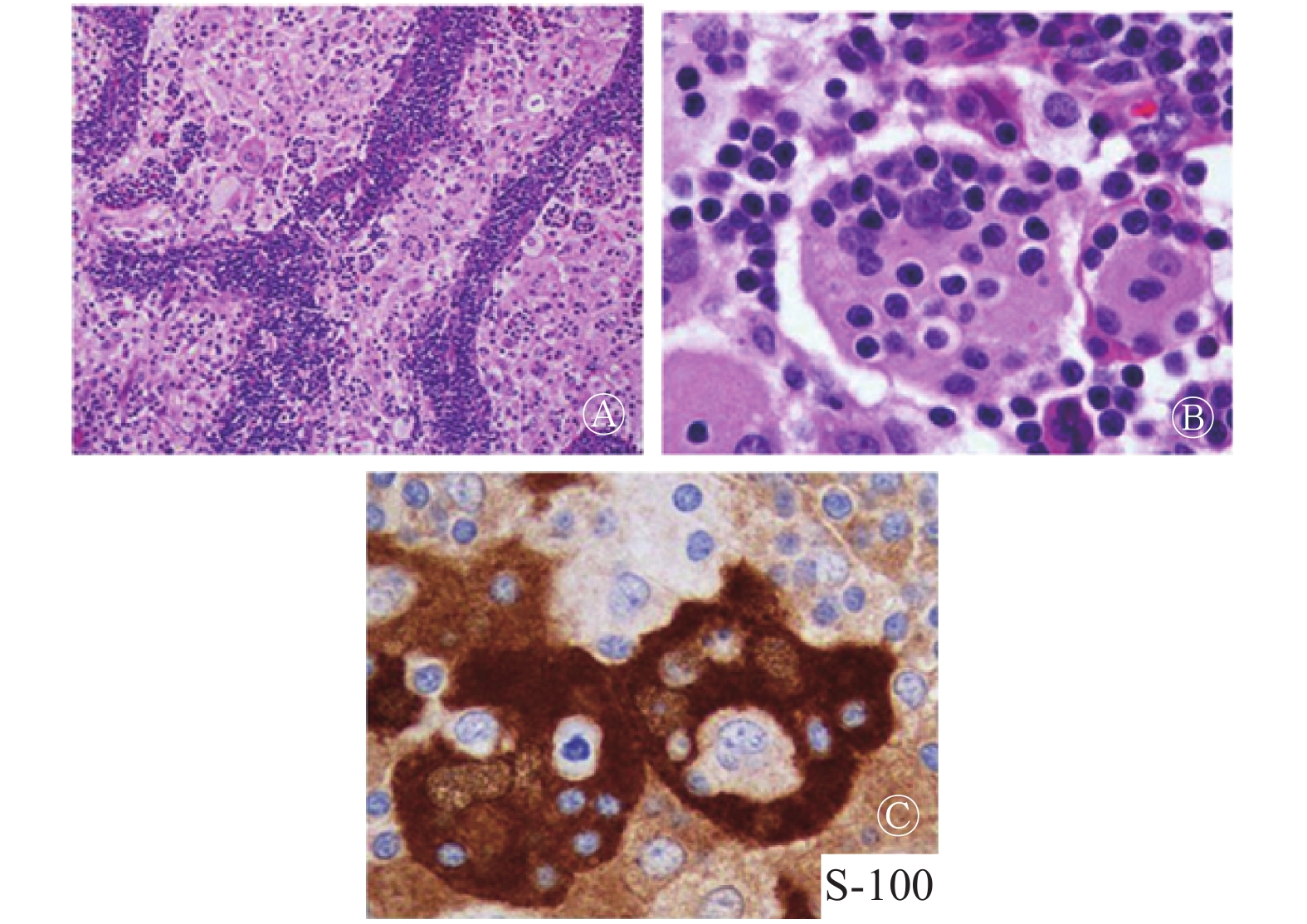
对右侧小腿行常规X射线检查未见明显异常,为明确诊断及病情分期行18F-FDG PET/CT检查,右侧胫骨上段见异常高代谢病灶,SUVmax=23.0,右侧胫骨骨皮质不规则变薄、毛糙,骨内膜不光滑、毛刷样改变。同机CT显示右侧胫骨髓腔内密度增高,与同水平18F-FDG代谢增高影范围一致,平均CT值约为34.6 HU(图1)。根据影像所见,18F-FDG PET/CT初步印象:骨淋巴瘤?不明原发灶所致骨髓转移?右侧胫骨活检病理学结果:纤维结缔组织中见大量合体细胞样组织细胞,胞质丰富淡染,细胞核染色质细腻,单核或双核,可见核仁,间质见大量浆细胞及中性粒细胞浸润和淋巴细胞浸润。免疫组织化学染色结果:组织细胞S-100蛋白(+)、组织细胞CD68 (散在+)、细胞增殖核抗原Ki67(约4%+)(图2)。

图 1 右胫骨Rosai-Dorfman病患者(女性,36岁)的18F-FDG PET/CT显像图
Figure 1. 18F-FDG PET/CT images of the patient with Rosai-Dorfman disease in right tibia (female, 36 years old)

图 2 右胫骨Rosai-Dorfman病患者(女性,36岁)的病理学免疫组化染色结果
Figure 2. Pathological immunohistochemistry staining images of Rosai-Dorfman disease in right tibia (female, 36 years old)
病理所见结合临床影像,初步诊断为RDD。确诊后患者接受一个疗程的放疗,恢复良好,症状明显减轻,目前仍在随访中。
-
RDD又称窦性组织细胞增生症,伴巨大淋巴结病变,是一种罕见的组织细胞增生性疾病,在世界卫生组织公布的肿瘤分类中被定义为原因不明的反应性病变,归于淋巴造血系统肿瘤[3];常表现为发作性加重和自发缓解。
RDD主要原发于淋巴结,表现为颈部无痛性淋巴结肿大,多伴发热、WBC增多、RBC沉降率增快、多克隆性丙种球蛋白血症和体重下降等,约43%的患者伴淋巴结外浸润病变[4]。此外,还有约25%的患者仅有淋巴结外病变而不伴淋巴结病,称为结外RDD,结外主要累及皮肤、眼睑、副鼻窦、软组织和骨骼,也有累及椎管的报道[5]。骨骼病变通常以溶骨性表现为主,不易与其他淋巴组织增生性疾病区分。耶鲁大学Foucar等[6]报道了一组较大规模的RDD病例(423例),大部分患者表现为淋巴结肿大,43%的患者合并结外浸润;其中骨受累患者约占7%,单纯孤立性骨骼浸润相当罕见,仅占0.7%。Mosheimer等[7]报道了一组108例RDD累及骨骼的患者,其中原发于骨的RDD占74.4%,最常见的症状是疼痛和局部肿胀,但大多数骨骼病灶为偶然发现。本例患者因间歇性疼痛就诊,并行全身18F-FDG PET/CT扫描,客观上为早期检出病变赢得了时机。
结外RDD特征性的病理学表现:低倍镜苏木精-伊红染色下可见病变组织内纤维组织增生和片状或不规则巢团状“浅染区”,周围混合有丰富的淋巴细胞、浆细胞、中性粒细胞浸润;高倍镜下可见体积大的多角形组织细胞、核大,可见小的嗜碱性核仁,有丰富的泡沫状胞质,胞质中可见“伸入现象”,即在胞质中吞噬有淋巴细胞和浆细胞,细胞异型性不明显,少见核分裂相。免疫组化结果示组织细胞表达S-100蛋白和CD68[8]。
原发于骨的RDD常规影像学表现无明显特征性,大多数病变表现为以骨髓腔为中心的溶骨性病变[9],边缘通常不规则,偶尔出现均匀一致的硬化性改变[10-11]。原发于长骨的病灶倾向于侵犯干骺端,如本例患者。在行PET/CT检查前常规CT表现为骨髓腔均匀稍高密度影,伴邻近骨质侵蚀,无钙化;RDD通常在MRI的T1加权序列中呈现与肌肉相近的等、低信号,反映出骨小梁间隙的正常骨髓被病变组织替换、浸润的过程,在液体翻转恢复序列T2加权像呈现明显的高信号[12]。PET/CT表现为18F-FDG代谢活跃病灶,其机制为纤维组织异常增生和组织细胞增生导致对葡萄糖的利用增加[13-14]。很多累及骨的病变可有类似表现,因此,原发于骨的RDD需要与很多相关疾病鉴别,包括骨髓炎、朗格汉斯细胞增生症、骨淋巴瘤、浆细胞瘤、原发性和转移性的骨肿瘤;由于大多数需鉴别的疾病也呈18F-FDG高摄取,因此,18F-FDG PET/CT区分此类疾病的价值有限。
18F-FDG PET/CT的优势:首先,它可以通过病灶部位18F-FDG代谢活性的增高检测到常规CT无法显示的病灶,还可检出体检无法触及的躯体深部的小病灶,早期发现处于亚临床期的骨髓病灶[15-16];其次,PET/CT通常是全身显像,相比常规放射学检查,可覆盖更多的部位,有利于发现更多的病灶,因此可修正常规放射学检查在疾病分期上的局限性,进行准确地系统分期;再次,通过18F-FDG PET/CT融合影像对病灶全身分布情况的显示,有利于筛选最适合的活检部位,提高活检的阳性率。RDD的确诊有赖于影像学检查、病理学检查和临床症状的相互结合与印证。
RDD大多为良性,临床进展较慢,但病变会因累及部位的不同而出现不同的临床症状。本研究结果显示,如果18F-FDG PET/CT显示以髓腔为中心的溶骨性破坏病变,临床伴有骨痛、发热等,同时伴有其他部位的多发代谢活跃的肿大淋巴结,除了要考虑骨髓炎、骨髓瘤、骨转移等常见疾病,还应当把有类似表现的罕见病(如RDD)列入鉴别诊断的范围之中。
利益冲突 本研究由署名作者按以下贡献声明独立开展,不涉及任何利益冲突。
作者贡献声明 王剑杰负责论文的起草、撰写和病理图片的收集与解析;马潞娜负责研究命题的提出;田智勇、陈旖文、刘学负责病例的收集、图像数据的获取;蒲朝煜负责论文的审核、最终版本的修订。
胫骨孤立性Rosai-Dorfman病18F-FDG PET/CT显像分析及文献复习
Appearance of a solitary Rosai-Dorfman disease of tibia on 18F-FDG PET/CT: A case report and literature review
-
摘要: Rosai-Dorfman病是一种罕见的组织细胞病,也称为窦性组织细胞增生症,伴有大量淋巴结肿大。典型表现为双侧颈部无痛淋巴结病,也可能发生淋巴结外性器官受累,包括皮肤、上呼吸道和骨骼。约8%的患者可发生骨受累。放射学检查对该病缺乏特异性,单纯依靠放射学检查很难确诊,病变的活检和病理检查是确诊的唯一途径。笔者报道了1例右腿间歇性疼痛的36岁Rosai-Dorfman病患者的18F-FDG PET/CT表现。该研究结果提示18F-FDG PET/CT融合显像在鉴别诊断、确定疾病的范围和指导活检取材方面有重要价值。Abstract: Rosai-Dorfman disease (RDD), also known as sinus histiocytosis with massive lymphadenopathy, is a rare histiocytic disease. Typically, a patient with RDD presents with painless bilateral cervical lymphadenopathy. The involvement of extranodal organs, such as the skin, upper respiratory tract, and bone, may also occur. Bone involvement occurs in approximately 8% of patients with RDD. Obtaining an affirmatory diagnosis by non-specific radiological findings is difficult. The final diagnosis can be achieved by biopsy and pathological examination of the lesion. The 18F-FDG PET/CT results of a young patient aged 36 with RDD who presented with intermittent pain in the right leg was reported in this article. This case illustrates the usefulness of functional fused modality in differentiating diagnosis and in identifying the extent of this disease and the appropriate sites for biopsy.
-
Key words:
-
图 1 右胫骨Rosai-Dorfman病患者(女性,36岁)的18F-FDG PET/CT显像图
Figure 1. 18F-FDG PET/CT images of the patient with Rosai-Dorfman disease in right tibia (female, 36 years old)
-
[1] Baker JC, Kyriakos M, McDonald DJ, et al. Primary Rosai-Dorfman disease of the femur[J]. Skeletal Radiol, 2017, 46(1): 129−135. DOI: 10.1007/s00256−016−2515−3. [2] Xu HC, Zhang F, Lu FZ, et al. Spinal Rosai-Dorfman disease: case report and literature review[J]. Eur Spine J, 2017, 26(S1): S117−127. DOI: 10.1007/s00586−017−4975−0. [3] 徐志锋, 潘爱珍, 李勤祥, 等. Rosai-Dorfman病的临床及影像表现[J]. 中华放射学杂志, 2018, 52(12): 936−940. DOI: 10.3760/cma.j.issn.1005−1201.2018.12.011.
Xu ZF, Pan AZ, Li QX, et al. The clinical and imaging characteristics of Rosai-Dorfman disease[J]. Chin J Radiol, 2018, 52(12): 936−940. DOI: 10.3760/cma.j.issn.1005−1201.2018.12.011.[4] Foucar E, Rosai J, Dorfman RF. Sinus Histiocytosis With Massive Lymphadenopathy: Current Status and Future Directions[J]. Arch Dermatol, 1998, 124(8): 1211−1214. DOI: 10.1001/archderm.1988.01670080023011. [5] 黄碧娟, 张盼, 陈自谦. 原发性椎管内Rosai-Dorfman病(附1例报告及文献复习)[J]. 医学影像学杂志, 2016, 26(12): 2189−2191.
Huang BJ, Zhang P, Chen ZQ. Primary rosai-dorfman disease (a case report and review of the literature)[J]. J Med Imaging, 2016, 26(12): 2189−2191.[6] Foucar E, Rosai J, Dorfman R. Sinus histiocytosis with massive lymphadenopathy (Rosai-Dorfman disease): review of the entity[J]. Semin Diagn Pathol, 1990, 7(1): 19−73. [7] Mosheimer BA, Oppl B, Zandieh S, et al. Bone Involvement in Rosai-Dorfman Disease (RDD): a Case Report and Systematic Literature Review[J]. Curr Rheumatol Rep, 2017, 19(5): 29. DOI: 10.1007/s11926−017−0656−6. [8] Alruwaii Z, Zhang Y, Larman T, et al. Rosai-Dorfman Disease of the Digestive System-Beware Vasculopathy: A Clinicopathologic Analysis[J]. Am J Surg Pathol, 2019, 43(12): 1644−1652. DOI: 10.1097/PAS.0000000000001343. [9] Tsang JS, Anthony MP, Wong MP, et al. The use of FDG-PET/CT in extranodal Rosai-Dorfman disease of bone[J]. Skeletal Radiol, 2012, 41(6): 715−717. DOI: 10.1007/s00256−012−1382−9. [10] 李勤祥, 潘爱珍, 赵海, 等. 骨骼Rosai-Dorfman病的影像学表现并文献复习(附2例报道)[J]. 国际放射医学核医学杂志, 2017, 41(6): 396−400. DOI: 10.3760/cma.j.issn.1673−4114.2017.06.004.
Li QX, Pan AZ, Zhao H, et al. Radiographic manifestations of skeleton Rosai-Dorfman disease: case report and review of related literature[J]. Int J Radiat Med Nucl Med, 2017, 41(6): 396−400. DOI: 10.3760/cma.j.issn.1673−4114.2017.06.004.[11] Gupta N, Verma R, Belho ES, et al. Isolated Extranodal Rosai-Dorfman Disease on 18F-FDG PET-CT Scan[J]. Indian J Nucl Med, 2019, 34(4): 319−320. DOI: 10.4103/ijnm.IJNM_152_19. [12] Rittner RE, Baumann U, Laenger F, et al. Whole-body diffusion-weighted MRI in a case of Rosai-Dorfman disease with exclusive multifocal skeletal involvement[J]. Skeletal Radiol, 2012, 41(6): 709−713. DOI: 10.1007/s00256−011−1328−7. [13] Zadeh MZ, Wen Z, States L, et al. An Isolated Osseous Rosai-Dorfman Disease Shown on FDG PET/CT[J]. Clin Nucl Med, 2019, 44(6): 485−488. DOI: 10.1097/RLU.0000000000002501. [14] Xu QG, Fu LP, Liu CY. Multimodality imaging-based evaluation of Rosai-Dorfman disease in the head and neck: A retrospective observational study[J]. Medicine, 2017, 96(51): e9372. DOI: 10.1097/MD.0000000000009372. [15] Shaikh F, Awan O, Mohiuddin S, et al. 18F-FDG PET/CT Imaging of Extranodal Rosai-Dorfman Disease with Hepatopancreatic Involvement-A Pictorial and Literature Review[J/OL]. Cureus, 2015, 7(12): e392[2019-05-09]. https://www.cureus.com/articles/3636−18f−fdg−petct−imaging−of−extranodal−rosai−dorfman−disease−with−hepatopancreatic−involvement−a−pictorial−and−literature−review. DOI: 10.7759/cureus.392. [16] Sher A, Orth R, McClain K, et al. PET/MR in the Assessment of Pediatric Histiocytoses: A Comparison to PET/CT[J]. Clin Nucl Med, 2017, 42(8): 582−588. DOI: 10.1097/RLU.0000000000001717. -

 点击查看大图

图(2)
计量
- 文章访问数: 2494
- HTML全文浏览量: 1677
- PDF下载量: 11