


-
功能不全性骨折(insufficiency fracture,IF)是应力性骨折的一种,也称为衰竭性骨折或矿物质缺乏性骨折,是正常或生理性肌肉活动作用于矿物质减少或弹性抵抗力减弱的骨骼而导致的脆性骨折。IF常见于绝经后骨质疏松的女性患者,也见于其他原因骨质疏松的患者,如长期服用激素、糖尿病、骨质软化及恶性肿瘤放疗后患者等[1-2]。IF患者常无临床症状或症状不明显,临床容易漏诊或误诊[3-5]。99Tcm-MDP SPECT全身骨显像对骨病的诊断具有较高的灵敏度,但特异度低,SPECT/CT可提高诊断的特异度,拓展了其临床应用范围。本研究回顾性分析90例骨质疏松患者的99Tcm-MDP SPECT全身骨显像及局部SPECT/CT融合显像的影像学资料,探讨联合显像对IF的诊断价值,以及对新鲜或陈旧性IF的鉴别诊断价值。
-
选取2012年12月至2017年6月于河南省骨科医院收治的符合骨质疏松症诊断标准的90例患者,其中男性32例、女性58例,年龄(67.56±7.68)岁。所有患者中,30~50岁的4例(男性2例、女性2例),50~60岁的2例(均为女性),60~70岁的45例(女性31例、男性14例),71~90岁的39例(女性23例、男性16例)。22例患者可疑IF,68例患者已确诊局部IF。
纳入标准:①符合骨质疏松诊断标准,采用双能X射线骨密度仪测量腰1~腰4椎体的骨密度,骨质疏松诊断标准采用世界卫生组织的诊断标准,即T值≤−2.5 SD[1];②无明显外伤史;③行SPECT全身骨显像及SPECT/CT融合显像检查,并于检查前签署知情同意书;④最终诊断为IF,具有典型的影像学特征并经6个月的影像学随访证实。排除标准:合并风湿及类风湿疾病、骨肿瘤和骨感染等患者。
-
显像仪器为德国西门子公司Siemens Symbia True point 型SPECT/CT仪。患者于扫描前3~4 h静脉注射99Tcm-MDP(江苏省原子医学研究所江原制药厂提供)740~1110 MBq。先行SPECT全身前位及后位平面骨显像,采集条件:能峰140 keV,窗宽20%,矩阵为256×1024,床速为20 cm/min。采集完成后患者保持体位不变,以全身骨显像检出的阳性部位为中心行局部SPECT/CT融合显像。SPECT显像条件:矩阵128×128,放大倍数1,360°采集,双探头各旋转180°,6°/帧,20 s/帧;CT扫描条件:管电压110 kV,管电流 50 mAs,层厚1.5 mm,螺距1。采用仪器自带软件融合SPECT/CT图像。
-
由3位具有10年以上阅片经验的核医学医师共同对 99Tcm-MDP SPECT全身骨显像、CT、SPECT/CT融合图像进行分析。99Tcm-MDP SPECT全身骨显像的阳性部位需排除显像剂摄取增高的关节部位。CT诊断骨折的标准:CT观察到骨折线、硬化带/线、椎体呈楔形或双凹变,可合并或不合并周围软组织肿胀。上述诊断需要排除溶骨性、成骨性、混合性骨质破坏和合并软组织肿块的病灶。SPECT/CT诊断新鲜骨折的标准:融合图像显示骨折且显像剂沿着骨折线两端摄取增高;骨质形态结构改变,但如无显像剂摄取为陈旧性骨折。IF的最终诊断需同时符合下述3条诊断标准:①无明确外伤史;②符合骨质疏松的诊断标准;③经过6个月以上的影像学随访证实为骨折。
-
99Tcm-MDP SPECT全身骨显像共检出病灶234个,均表现为不同程度的点状、条形或团状显像剂摄取增高;局部SPECT/CT显像可见骨折线、硬化带/线或椎体高度降低,显像剂沿着骨折线周围摄取增高,提示均为IF且随访证实均为真阳性。99Tcm-MDP SPECT全身骨显像联合SPECT/CT融合显像对骨质疏松患者IF的诊断灵敏度为100%(234/234),准确率为100%。90例患者中,单发IF 21例、多发IF 69例,合计234个IF病灶,具体部位见表1。
IF部位 例数(例) 病灶数(个) 骨盆 22 78 腰椎 36 49 胸椎 24 46 肋骨 11 45 股骨 6 8 胫骨 4 4 跟骨 2 3 胸骨 1 1 合计 234 注:表中,IF:功能不全性骨折 表 1 90例骨质疏松患者IF的分布部位及病灶数
Table 1. Distribution of insufficiency fracture and the number of lesions in 90 cases of osteoporosis
68.89%(62/90)的患者联合显像检出的IF病灶数目多于显像之前已知的病灶数目。22例患者的解剖影像学结果为阴性,而联合显像为阳性;68例患者行联合显像之前,局部解剖影像学检出患者局部骨骼有IF,显像之后,40例患者的IF病灶部位多于显像之前诊断的病灶数目。234个IF病灶,其中86个IF病灶患者的临床症状不明显,由联合显像首先发现(图1、图2)。
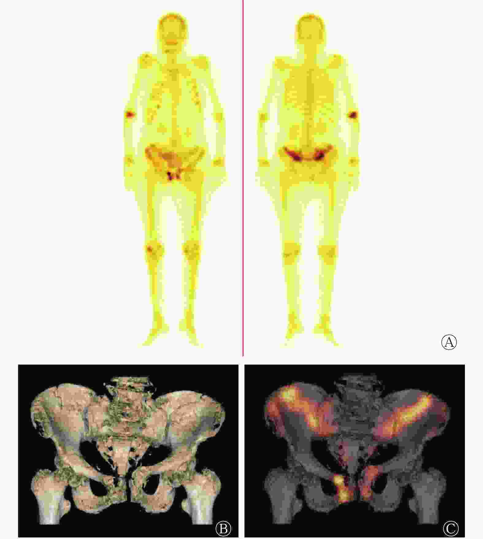
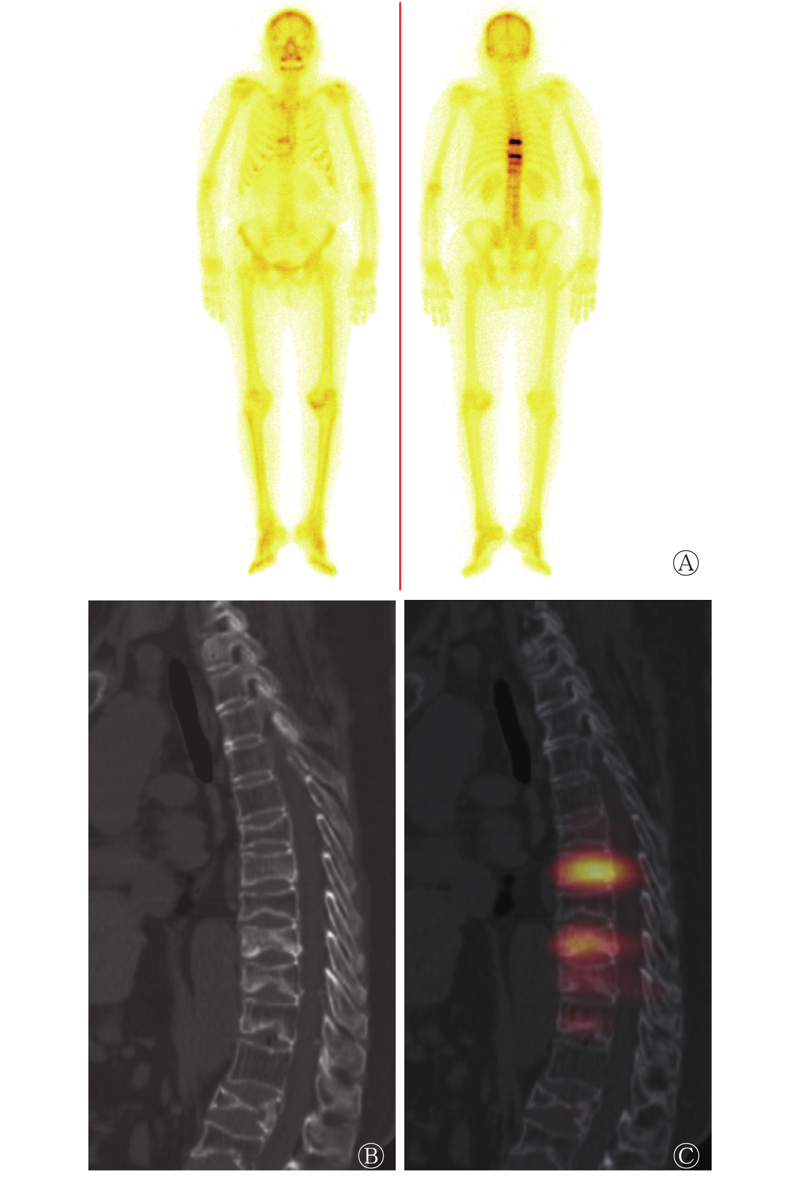
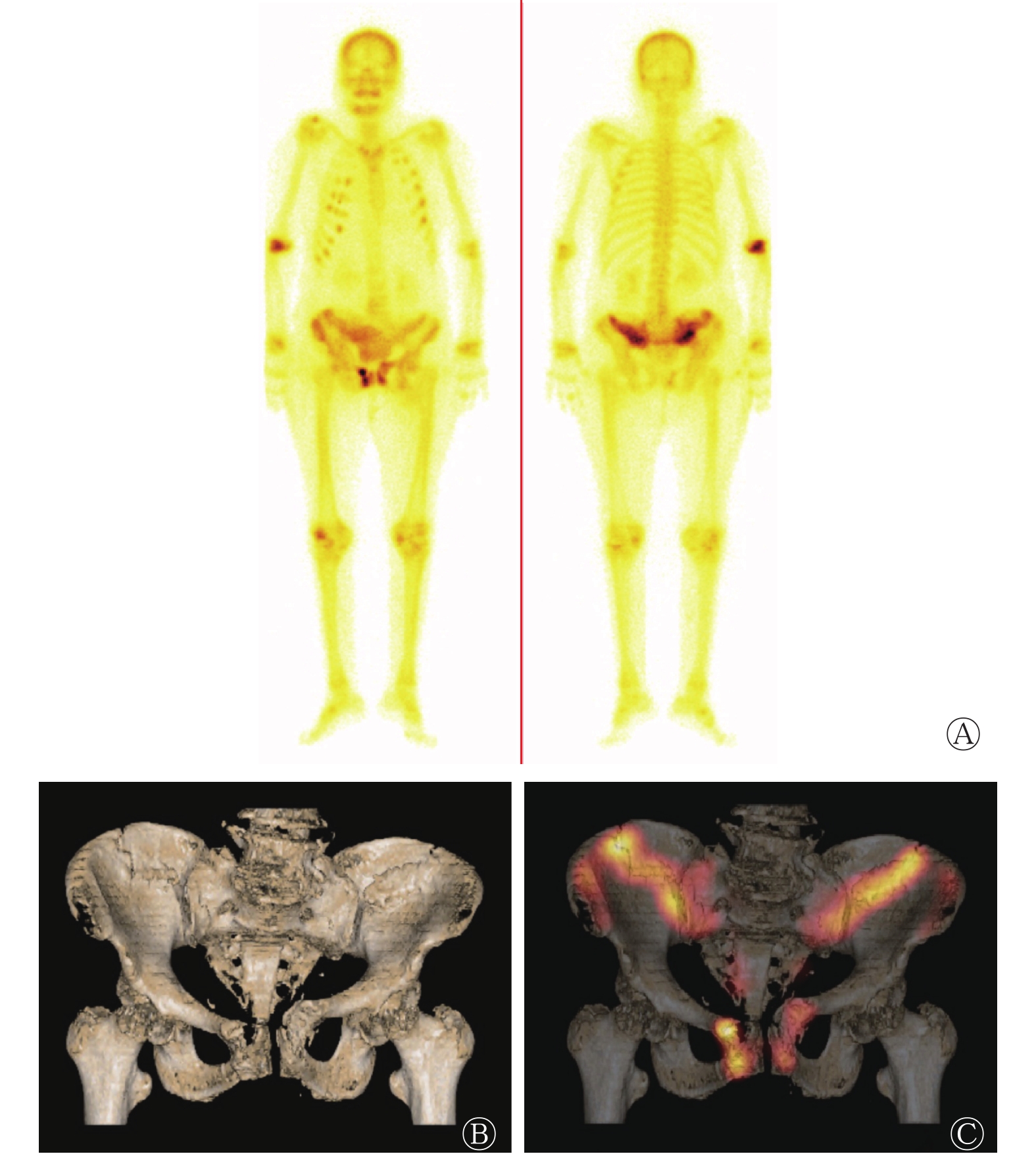
图 1 全身多发功能不全性骨折患者(女性,76岁)的99Tcm-MDP SPECT全身骨显像图(A)和骨盆SPECT/CT显像图(B、C) 患者臀部疼痛不适15 d。图中,A:双侧多发肋骨骨折呈“串珠状”改变,骶椎、双侧髂骨及双侧耻骨显像剂局部摄取增高;B:CT图像示骨盆呈骨质疏松改变,骶椎双侧、双侧髂骨翼及双侧耻骨骨折;C:融合图像示显像剂沿着骨折线摄取增高。MDP:亚甲基二膦酸盐;SPECT:单光子发射计算机体层摄影术;CT:计算机体层摄影术
Figure 1. 99Tcm-MDP SPECT whole-body bone scan (A) and local SPECT/CT tomography (B, C) of a 76-year-female patient with multiple insufficiency fracture
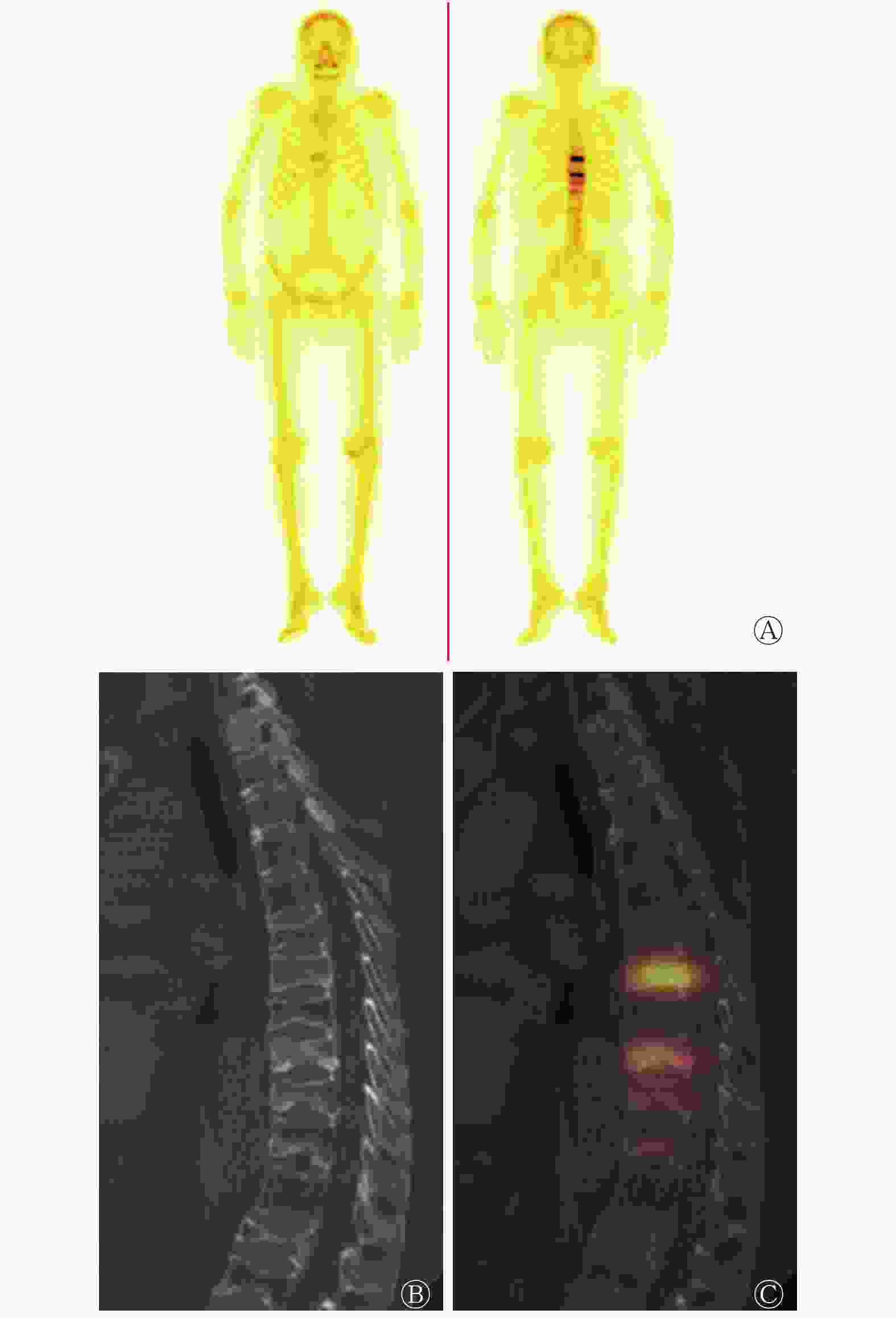
图 2 全身多发功能不全性骨折患者(女性,69岁)的99Tcm-MDP SPECT全身骨显像图(A)和腰椎SPECT/CT显像图(B、C) 患者胸背部疼痛不适10余天。图中,A:胸9、胸11椎体显像剂条形摄取增高;B:CT图像示胸8~胸12、腰1、腰3~腰4椎体高度不同程度降低;C:融合图像示胸9、胸11椎体显像剂呈条形摄取增高,提示新鲜骨折,余形态改变的椎体显像剂无明显摄取,提示陈旧性骨折。MDP:亚甲基二膦酸盐;SPECT:单光子发射计算机体层摄影术;CT:计算机体层摄影术
Figure 2. 99Tcm-MDP SPECT whole-body bone scan (A) and local SPECT/CT tomography (B, C) of a 69-year-female patient with multiple insufficiency fracture
在局部SPECT/CT融合显像检查范围内,CT显示骨折病灶293个,其中234个显像剂摄取明显增高,提示为新鲜IF;59处无明显显像剂摄取或略显显像剂摄取,提示为陈旧性IF,随访证实均为陈旧性IF(图2)。SPECT/CT对新鲜IF的诊断灵敏度为100%(234/234)、特异度为100%(59/59)。
99Tcm-MDP SPECT全身骨显像显示11例患者的肋骨IF,其中9例患者的肋骨IF呈点状纵向排列的显像剂摄取增高(图1)。在22例骨盆IF患者中,6例骶椎显像剂摄取增高,呈“H”形改变(图3),占总数的27.28%。
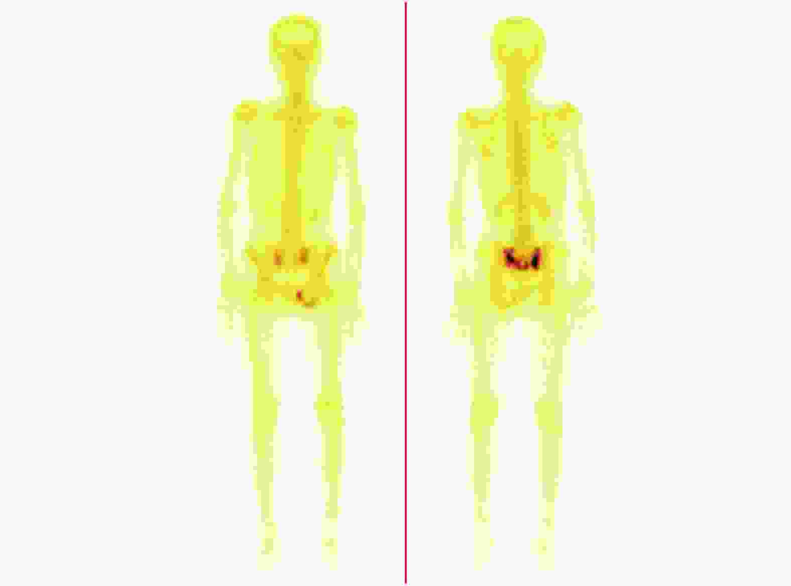
图 3 骨盆多发功能不全性骨折患者(女性,71岁)的99Tcm-MDP SPECT全身骨显像图
Figure 3. 99Tcm-MDP SPECT whole-body bone scan of a 71-year-female patient with pelvic multiple insufficiency fracture
-
人体骨骼承受不了正常的日常活动而造成的骨折称为IF,易发生于身体承重区或易受到扭曲作用力的部位,如胸腰椎、骨盆及自由下肢骨等。IF常见于老年人或身体衰弱的骨质疏松患者,多无明显外伤史,因临床症状不明显而容易漏诊。同时由于骨骼活动的整体性及力学传导,如脊柱、骨盆的日常活动均是多关节、多部位同时运动,因此IF多为多发骨折,很少单独一处骨折[6-9]。数字化摄影(DR)是骨关节疾病首选的检查方法,但其对解剖结构重叠部位的病灶显像欠佳。MRI属于断层影像学检查方法,不受解剖结构重叠的影响,且对隐匿性骨折的显示具有较高的灵敏度。但MRI检查费用较高,不适宜对骨质疏松患者行全身IF筛查;同时对装有心脏起搏器等顺磁性物质的患者禁行MRI检查[10-11]。本研究的90例患者中,多发IF患者有69例,占76.67%;常见部位是骨盆、腰椎、胸椎和肋骨;共有234处病灶,其中86处病灶患者的临床症状不明显,由联合显像首先发现。本研究结果进一步证实,骨质疏松患者的IF有一定的好发部位、容易多发且临床症状可能会不明显。
IF发生后6~72 h即发生一系列病理、生理变化,骨折部位的成骨细胞代谢活跃,修复活动增强以及软组织出血等,致使骨显像剂异常浓聚于骨折处,在图像上呈现为“热区”。因此,SPECT全身骨显像对IF的早期诊断具有较高的价值,灵敏度近100%,可谓“金标准”,全身骨显像为一次性成像,可发现全身骨骼病变,能准确、全面评估病灶累及范围[12-13]。本研究中90例骨质疏松患者的234个IF病灶,全身骨显像均表现为显像剂摄取增高,但全身骨显像对疾病诊断的特异度较低,需要与肿瘤、感染和退变等疾病进行鉴别。有研究显示,骶椎多发骨折呈“H”形或蝴蝶形改变[10],但也有研究显示并不是所有骶椎的IF均表现为“H”形,转移瘤也可表现为此征象[14]。本研究中22例骨盆IF患者,其中仅6例骶椎显像剂摄取增高,呈“H”形改变。CT可弥补全身骨显像特异度低的缺点,SPECT/CT融合显像可对病灶的解剖结构和功能信息进行全面显示,提高疾病诊断的特异度[15-16]。本研究中全身骨显像的234个阳性病灶,经SPECT/CT融合显像之后,可观察到骨折线、硬化线和椎体形态改变,提高了IF的诊断特异度。
对疼痛责任病灶的确定,即对陈旧性或新鲜IF的鉴别有重要的临床意义。CT只能从形态学上观察骨质结构的改变,不能准确区分新鲜IF或陈旧性IF。SPECT/CT则能弥补上述不足,IF急性期表现为病灶对显像剂摄取明显增高,随着IF的修复,显像剂摄取程度逐渐降低,这提示可依据显像剂摄取程度判断大概的骨折时间。如骨折端显像剂摄取明显增高,提示为新鲜IF;如显像剂摄取不明显,则提示陈旧性IF。本研究中CT检查范围内显示骨折病灶293个,其中234个病灶对显像剂的摄取明显增高,随访证实SPECT/CT对新鲜IF的诊断灵敏度为100%,可准确鉴别新鲜或陈旧性IF。但骨折修复是一个复杂的病理过程,不同年龄、性别、骨折部位和个体的营养状况均会影响到骨折的愈合过程,是否可依据骨显像剂摄取程度精确判断骨折时间,还需进一步研究。
总之,SPECT全身骨显像一次成像可对全身骨骼进行评估,联合局部SPECT/CT融合显像可准确诊断IF,并实现对新鲜或陈旧IF的鉴别诊断,值得临床推广。
利益冲突 本研究由署名作者按以下贡献声明独立开展,不涉及任何利益冲突。
作者贡献声明 张斌青负责方法的建立、资料的获取和论文的撰写;张国庆、刘玉珂负责图像和数据的分析;郭会利负责方法的设计和论文的审阅。
全身骨显像联合局部SPECT/CT对功能不全性骨折的诊断价值
Diagnostic value of whole-body bone scan and local SPECT/CT for insufficiency fracture
-
摘要:
目的 探讨99Tcm-MDP SPECT全身骨显像联合局部SPECT/CT融合显像对骨质疏松患者功能不全性骨折(IF)的诊断价值。 方法 回顾性分析2012年12月至2017年6月于河南省骨科医院收治的90例符合骨质疏松症诊断标准的患者的99Tcm-MDP SPECT全身骨显像和阳性部位的局部SPECT/CT融合显像结果,分析联合显像对IF的临床诊断效能。90例患者中,男性32例、女性58例,年龄(67.56±7.68)岁,其中22例可疑IF、68例已确诊局部IF。所有患者的最终诊断结果均经过6个月以上临床及影像学随访证实。 结果 99Tcm-MDP SPECT全身骨显像共检出IF病灶234个,均表现为显像剂摄取增高,病变部位及个数为骨盆78个、腰椎49个、胸椎46个、肋骨45个、股骨8个、胫骨4个、跟骨3个及胸骨1个。全身骨显像联合局部SPECT/CT融合显像对IF的诊断灵敏度为100%(234/234)。68.89%(62/90)的患者联合显像检出的IF数目多于显像之前已知的骨折数目。在局部SPECT/CT融合显像检查范围内,CT显示骨折病灶293个,其中SPECT/CT诊断新鲜IF病灶234个、陈旧性IF病灶59个,SPECT/CT对新鲜IF的诊断灵敏度为100%(234/234)。 结论 99Tcm-MDP SPECT全身骨显像联合局部SPECT/CT融合显像对骨质疏松患者IF病灶数目的全身显示及诊断具有较高的临床应用价值。 -
关键词:
- 骨折,应力性 /
- 体层摄影术,发射型计算机,单光子 /
- 体层摄影术,X线计算机 /
- 99m锝美罗酸盐
Abstract:Objective To investigate the diagnostic value of 99Tcm-MDP SPECT whole-body bone scan and local SPECT/CT tomography fusion imaging for insufficiency fracture (IF). Methods Ninety patients (including 32 males and 58 females, aged 67.56±7.68, 22 cases with suspected IF, 68 cases with confirmed local IF) who were either diagnosed with IF in their partial skeleton or suspected to suffer from IF were enrolled in this study. The diagnostic value of 99Tcm-MDP SPECT whole-body bone scan and local SPECT/CT tomography fusion imaging for IF were retrospectively analyzed. The correct diagnosis were confirmed by clinical assessment and over 6 months of imaging follow-up. Results A total of 234 lesions were detected in 90 patients by 99Tcm-MDP SPECT whole-body bone scan, which showed high radioactivity in all lesions. The common sites of the lesions were the pelvis (78 lesions), lumbar vertebrae (49 lesions), thoracic vertebrae (46 lesions), ribs (45 lesions), femur (8 lesions), tibia (4 lesions), calcaneus (3 lesions), and sternus (1 lesion). The sensitivity of 99Tcm-MDP whole-body bone scan and local SPECT/CT tomography fusion imaging for IF was 100% (234/234). A greater number of lesions were determined in approximately 68.89% (62/90) of the patients by combined imaging than the lesions which were confirmed as IF before combined imaging. A total of 293 fractures were identified in the scope of CT examination; of these, 234 were diagnosed as acute fractures and 59 were diagnosed as chronic fractures by SPECT/CT. The diagnostic sensitivity of SPECT/CT toward acute IF was 100% (234/234). Conclusion 99Tcm-MDP SPECT whole body-bone scan and local SPECT/CT fusion imaging has high clinical value in the systemic display and diagnosis of IF in patients with osteoporosis. -
图 1 全身多发功能不全性骨折患者(女性,76岁)的99Tcm-MDP SPECT全身骨显像图(A)和骨盆SPECT/CT显像图(B、C) 患者臀部疼痛不适15 d。图中,A:双侧多发肋骨骨折呈“串珠状”改变,骶椎、双侧髂骨及双侧耻骨显像剂局部摄取增高;B:CT图像示骨盆呈骨质疏松改变,骶椎双侧、双侧髂骨翼及双侧耻骨骨折;C:融合图像示显像剂沿着骨折线摄取增高。MDP:亚甲基二膦酸盐;SPECT:单光子发射计算机体层摄影术;CT:计算机体层摄影术
Figure 1. 99Tcm-MDP SPECT whole-body bone scan (A) and local SPECT/CT tomography (B, C) of a 76-year-female patient with multiple insufficiency fracture
图 2 全身多发功能不全性骨折患者(女性,69岁)的99Tcm-MDP SPECT全身骨显像图(A)和腰椎SPECT/CT显像图(B、C) 患者胸背部疼痛不适10余天。图中,A:胸9、胸11椎体显像剂条形摄取增高;B:CT图像示胸8~胸12、腰1、腰3~腰4椎体高度不同程度降低;C:融合图像示胸9、胸11椎体显像剂呈条形摄取增高,提示新鲜骨折,余形态改变的椎体显像剂无明显摄取,提示陈旧性骨折。MDP:亚甲基二膦酸盐;SPECT:单光子发射计算机体层摄影术;CT:计算机体层摄影术
Figure 2. 99Tcm-MDP SPECT whole-body bone scan (A) and local SPECT/CT tomography (B, C) of a 69-year-female patient with multiple insufficiency fracture
图 3 骨盆多发功能不全性骨折患者(女性,71岁)的99Tcm-MDP SPECT全身骨显像图
Figure 3. 99Tcm-MDP SPECT whole-body bone scan of a 71-year-female patient with pelvic multiple insufficiency fracture
表 1 90例骨质疏松患者IF的分布部位及病灶数
Table 1. Distribution of insufficiency fracture and the number of lesions in 90 cases of osteoporosis
IF部位 例数(例) 病灶数(个) 骨盆 22 78 腰椎 36 49 胸椎 24 46 肋骨 11 45 股骨 6 8 胫骨 4 4 跟骨 2 3 胸骨 1 1 合计 234 注:表中,IF:功能不全性骨折  下载: 导出CSV
-
[1] 黄公怡. 骨质疏松性骨折及其临床特点[J]. 中华骨科杂志, 2008, 28(1): 74−77. DOI: 10.3321/j.issn:0253-2352.2008.01.017.
Huang GY. Osteoporotic fracture and its clinical characteristics[J]. Chin J Orthop, 2008, 28(1): 74−77. DOI: 10.3321/j.issn:0253-2352.2008.01.017.[2] Torres PAU, Cohen-Solal M. Evaluation of fracture risk in chronic kidney disease[J]. J Nephrol, 2017, 30(5): 653−661. DOI: 10.1007/s40620-017-0398-6. [3] 赵永杰, 谭伟光, 付至江, 等. 衰竭骨折11例的临床确诊困难原因分析[J]. 实用医学杂志, 2017, 33(23): 3963-3965. DOI: 10.3969/j.issn.1006-5725.2017.23.029.
Zhao YJ, Tan WG, Fu ZJ, et al. Analysis of the causes of difficulties in clinical diagnosis of 11 failure fractures[J]. J Pract Med, 2017, 33(23): 3963-3965. DOI: 10.3969/j.issn.1006-5725.2017.23.029.[4] Lapina O, Tiškevičius S. Sacral insufficiency fracture after pelvic radiotherapy: adiagnostic challenge for a radiologist[J]. Medicina, 2014, 50(4): 249−254. DOI: 10.1016/j.medici.2014.09.006. [5] 付占立, 龙蓉, 刘萌, 等. 骶骨衰竭性骨折全身骨显像误诊为骨转移一例[J]. 中华核医学与分子影像杂志, 2016, 36(4): 359−360. DOI:10.3760/cma.j.issn.2095-2848.2016.04.019.
Fu ZL, Long R, Liu M, et al. A case of sacral insufficiency fracture misdiagnosed as bone metastases on whole body bone scan[J]. Chin J Nucl Med Mol Imaging, 2016, 36(4): 359−360. DOI: 10.3760/cma.j.issn.2095-2848.2016.04.019.[6] 刘仁爵, 周映华. 肋骨衰竭性应力骨折[J]. 实用放射学杂志, 2000, 16(5): 309-311. DOI: 10.3969/j.issn.1002-1671.2000.05.018.
Liu RJ, Zhou YH. Stress fracture of rib caused by failure[J]. J Pract Radiol, 2000, 16(5): 309-311. DOI:10.3969/j.issn.1002-1671.2000.05.018.[7] Wagner D, Hofmann A, Kamer L, et al. Fragility fractures of the sacrum occur in elderly patients with severe loss of sacral bone mass[J]. Arch Orthop Trauma Surg, 2018, 138(7): 971−977. DOI: 10.1007/s00402-018-2938-5. [8] Al-faham Z, Rydberg JN, Oliver Wong CY. Use of SPECT/CT with 99mTc-MDP bone scintigraphy to diagnose sacral insufficiency fracture[J]. J Nucl Med Technol, 2014, 42(3): 240−241. DOI: 10.2967/jnmt.114.137547. [9] 丁浩源, 陈跃, 朱艳, 等. 功能不全性骨折的全身骨显像及局部SPECT/CT断层显像特点[J]. 中国医学影像学杂志, 2017, 25(11): 858−861. DOI: 10.3969/j.issn.1005-5185.2017.11.015.
Ding HY, Chen Y, Zhu Y, et al. Features of whole-body bone scan and local SPECT/CT tomography of insufficiency fracture[J]. Chin J Med Imaging, 2017, 25(11): 858−861. DOI: 10.3969/j.issn.1005-5185.2017.11.015.[10] 柴晓媛, 陈光强, 龚建平, 等. 以边缘硬化和破坏同时存在的骨盆多发衰竭性骨折[J]. 中华医学杂志, 2014, 94(5): 393. DOI: 10.3760/cma.j.issn.0376-2491.2014.05.022.
Chai XY, Chen GQ, Gong JP, et al. Multiple failure fractures of the pelvis with both marginal hardening and destruction[J]. Natl Med J China, 2014, 94(5): 393. DOI: 10.3760/cma.j.issn.0376-2491.2014.05.022.[11] Krestan C, Hojreh A. Imaging of insufficiency fractures[J]. Eur J Radiol, 2009, 71(3): 398−405. DOI: 10.1016/j.ejrad.2008.04.059. [12] 张斌青, 宋青凤, 刘玉珂, 等. 99Tcm-MDP SPECT/CT图像融合诊断隐匿性骨折[J]. 中国医学影像技术, 2014, 30(4): 587−590.
Zhang BQ, Song QF, Liu YK, et al. 99Tcm-MDP SPECT/CT fusion in diagnosis of occult bone fractures[J]. Chin J Med Imaging Technol, 2014, 30(4): 587−590.[13] 王妮, 王喆, 李成, 等. 全身骨显像及局部SPECT/CT断层显像在疲劳性骨折诊断中的临床价值[J]. 中华核医学与分子影像杂志, 2016, 36(3): 261−262. DOI: 10.3760/cma.j.issn.2095-2848.2016.03.013.
Wang N, Wang Z, Li C, et al. Diagnostic value of whole-body bone scan and SPECT/CT for stress fracture[J]. Chin J Nucl Med Mol Imaging, 2016, 36(3): 261−262. DOI: 10.3760/cma.j.issn.2095-2848.2016.03.013.[14] Fujii M, Abe K, Hayashi K, et al. Honda sign and variants in patients suspected of having a sacral insufficiency fracture[J]. Clin Nucl Med, 2005, 30(3): 165−169. DOI: 10.1097/00003072-200503000-00004. [15] 李艳梅, 杨吉琴, 王莹, 等. 全身骨显像联合 SPECT/CT显像对骨纤维异常增殖症的诊断价值[J]. 中华核医学与分子影像杂志, 2015, 35(6): 470−473. DOI: 10.3760/cma.j.issn.2095-2848.2015.06.012.
Li YM, Yang JQ, Wang Y, et al. Diagnostic value of whole-body bone scan combined with SPECT/CT imaging for osteofibrous dys-plasia[J]. Chin J Nucl Med Mol Imaging, 2015, 35(6): 470−473. DOI: 10.3760/cma.j.issn.2095-2848.2015.06.012.[16] 张斌青, 刘云, 张敏, 等. SPECT/CT融合显像对腰椎峡部裂的诊断增益价值[J]. 中国临床医学影像杂志, 2017, 28(3): 216−219. DOI: 10.3969/j.issn.1008-1062.2017.03.017.
Zhang BQ, Liu Y, Zhang M, et al. The additional value of 99Tcm-MDP SPECT/CT for diagnosing lumbar spondylolysis[J]. J Chin Clin Med Imaging, 2017, 28(3): 216−219. DOI: 10.3969/j.issn.1008-1062.2017.03.017. -

 点击查看大图
计量
- 文章访问数: 3402
- HTML全文浏览量: 2227
- PDF下载量: 12